Geometric Optics of thick lenses and Matrix methods - PowerPoint PPT Presentation
1 / 27
Title:
Geometric Optics of thick lenses and Matrix methods
Description:
Geometric Optics of thick lenses and Matrix methods Mathematical treatment of refraction for thick lenses shows the existence of principal planes in the paraxial ... – PowerPoint PPT presentation
Number of Views:586
Avg rating:3.0/5.0
Title: Geometric Optics of thick lenses and Matrix methods
1
Geometric Optics of thick lenses and Matrix
methods
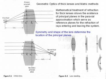
Mathematical treatment of refraction for thick
lenses shows the existence of principal planes in
the paraxial approximation which serve as
reference planes for the refraction of rays
entering and leaving the system.
Symmetry and shape of the lens determine the
location of the principal planes.
2
After an analytical treatment1 of refraction for
a thick lens geometry
Rough approximation for ordinary glass lenses in
air
1Complete derivation can be found in Morgan,
Introduction to Geometrical and Physical Optics.
Also, the Newtonian form holds
Fig. 6.4 Thick-Lens geometry
3
Note that as dl ? 0, this will yield the thin
lens result. Convention h1, h2 gt 0 then H1, H2
is to the right of V1,V2, and conversely if h1,
h2 lt 0 then H1, H2 is to the left of V1,V2 Again,
H1 and H2 refer to axial points through the
principal planes.
Now, consider a compound lens consisting of two
thick lenses L1 and L2, with the usual parameters
so1, si1, f1 and so2, si2 and f2, as shown on the
next slide.
si, so are image and object distances for the
combination as a whole and are measured with
respect to H1 and H2.
Note that the sign is important and distances gt 0
indicate that H1 or H2 are to the right of H11 or
H22.
4
Equivalent thick lens representation of a
compound lens
Fig. 6.5 A compound thick lens.
Note that if the lenses are thin, the pairs of
points H11, H12 and H21, H22 coalesce into a
single point and d becomes the center to center
lens separation.
5
Consider the example of a compound thin lens
below (individual lenses are thin). f1 -30 cm,
f2 20 cm, d 10 cm. Then, the effective focal
length is
Since these are thin lenses the principal planes
converge to single points O1, O2
A compound thin lens
6
Analytical Ray Tracing
3D
2D
Example of a computer program for ray tracing.
7
Consider a ray tracing analysis using the
paraxial approximation, sin? ? ? At point P1
Fig. 6.7 Ray Geometry for thick lenses
8
D1 is called the power of a single refracting
surface. For a thin lens,
This is done for cosmetic reasons.
Also, from the geometry
Thus, in matrix form we can write
9
Development of Matrix Method
Introduce ray vectors 2 ? 1 column matrix and a
2 ? 2 refraction matrix
Thus, we can define a 2 ? 2 transfer matrix
ray at P2 ray at P1
10
Continuing with the second interface in the
figure (Fig. 6.7)
Note that the determinant must be 1 and is a
check of the system matrix. After multiplying out
the system matrix, its components can be written
explicitly
Where d21 dl is the lens thickness and the
refractive index of the lens is nt1 nl.
11
Note that an examination of the system matrix A
gives
The lens is taken to be in air, as represented by
the powers D1 and D2.
We observe that this is just the reciprocal of
the focal length of a thick lens such that
a121/f , and the lens power is 1/f . More
generally, if the media are different on both
sides we would have
Thus, the matrix method involving 2 ? 2
refraction and transfer matrices enables a
determination of fundamental optical system
parameters such as the system focal lengths and
position of both principal planes relative to the
lens vertices.
12
image ray
object ray
The first operator T10 transfers the reference
point from the object (i.e., PO to P1). The next
operator A21 then carriers the ray through the
lens. A final transfer operator TI2 brings it to
the image plane, PI.
T transfer matrix R refraction matrix A
system matrix
13
Example of a complex lens system analyzed with
the Matrix Method
The result of the matrix method easily allows for
the solution of the basic lens parameters such as
the focal length and position of the principal
planes relative to the vertices of the outer
lenses.
Fig. 6.10 A Tessar lens system.
14
As another example, consider a system of thin
lenses in which dl ? 0. Note that the power of a
thin lens is D1 D2 D. Then
Suppose that two thin lenses are separated by
distance d
Then, the system matrix can be written as
15
Remember that
Where ni1 nt2 1 and V1 O1 and V2 O2 for
a thin lens
Note that the locations of the principal planes
H1 and H2 strongly depend on d, which can affect
on which side of the lenses the planes are
located. It is worth noting that a lens system
composed of N thin lenses can easily be treated
in the same manner for calculating the focal
lengths and locations of the principal planes.
16
A similar analysis can be performed for a mirror
in the paraxial approximation. The result is
Note that for a plane mirror R ? ? and the system
matrix for a mirror reduces to
17
Lens Aberrations Deviations from the
corresponding paraxial approximation Chromatic
Aberrations n(?) and ray components having
different colors have different effective focal
lengths Monochromatic Aberrations Spherical
aberration, coma, astigmatism Recall that we used
sin ? ? ? (first order theory in the paraxial
approx.) Including addition terms in sin ? ? ? -
? 3/3! leads to the third-order theory which can
explain the monochromatic aberrations. Remember
that for a single refracting spherical interface
in the 1st order approx
If the approximation for the OPL (lo li) are
improved, the 3rd order treatment gives
Where h is the distance above the optical axis as
shown in the figure.
18
Rays striking the surface at a greater distance
(marginal rays) are focused closer to the vertex
V than are the paraxial rays and creates
spherical aberration.
19
Marginal rays are bent too much and focused in
front of paraxial rays. Distance between the
intersection of marginal rays and the paraxial
focus, Fi, is known as the L?SA (longitudinal
spherical aberration). Note SA is positive for
convex lens and negative for a concave lens. T?SA
(transverse SA) is the transverse deviation
between the marginal and paraxial rays on a
screen placed at Fi. If the screen is moved to
the position ?LC the image blur will have its
smallest diameter, known as the circle of least
confusion, which is the best place to observe
the image.
20
Rule of thumb Incident ray will undergo a
minimum deviation when ?i ? ?T. Remember the
dispersing prism
Note that a planar-convex lens can be
approximated as two prisms.
?i
?1T
?2T gt ?1T and the lower prism results in a
greater deviation.
?2T
For an object at ?, the round side of lens facing
the object will suffer a minimum amount of SA.
Fig. 6.16 Spherical Aberration for a
planar-convex lens in both orientations.
21
Similarly if the object and image are to be
nearly equidistant from the lenses (so si 2f),
an Equi-Convex shaped lens minimizes SA.
Marginal rays give smaller image ? negative coma
Coma (comatic aberration) is associated with the
fact that the principle planes are really curved
surfaces resulting in a different MT for both
marginal and central rays. Since MT -si/so ,
the curved nature of the principal surface will
result in different effective object and image
distances, resulting in different transverse
magnifications. The variation in MT also depends
on the location of the object which can result in
a negative (a) or positive coma (b) and (c), as
demonstrated in the left figure.
Marginal rays give larger image ? positive coma
22
The imaging of a point at S can result in a
comet-like tail, known as a coma flare and
forms a comatic circle on the screen ?
(positive coma in this case). This is often
considered the worst out of all the aberrations,
primarily because of its asymmetric configuration.
23
Astigmatism The Meridional Plane contains the
chief ray which passes through the center of the
aperture and the optical axis. The Sagittal Plane
contains the chief ray and is perpendicular to
the meridional plane. Fermats principle shows
that planes containing the tilted rays will give
a shorter focal length, which depends on the (i)
power of the lens and the (ii) angle of
inclination. The result is that there is both a
meridional focus FT and a sagittal focus FS.
Tilted rays have a shorter focal length.
24
Astigmatism Note that the cross-section of the
beam changes from a circle (1) ? ellipse (2) ?
line (primary image 3)? ellipse (4) ? circle of
least confusion (5) ? ellipse (6) ? line
(secondary image 7) .
Focal length difference FS-FT depends on power D
of lens and angle of rays.
25
Chromatic Aberrations Since the index depends on
the wavelength then we can expect that the focal
length will depend on the wavelength.
f
?
26
Circle of least confusion
27
(No Transcript)

