Explain how sonar and ultrasound imaging work - PowerPoint PPT Presentation
Title:
Explain how sonar and ultrasound imaging work
Description:
RECOGNIZE WHAT FACTORS AFFECT THE SPEED OF SOUND MEDIUM-rubber slows vibrations, used as soundproofing 2. Temperature-higher the temperature, faster the sound – PowerPoint PPT presentation
Number of Views:90
Avg rating:3.0/5.0
Title: Explain how sonar and ultrasound imaging work
1
Explain how sonar and ultrasound imaging work
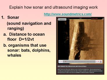
- http//www.soundmetrics.com/
- Sonar
- (sound navigation and ranging)
- a. Distance to ocean floor D1/2vt
- b. organisms that use sonar bats, dolphins,
whales
2
ULTRASOUND IMAGING
- SONOGRAMS
- frequency of 1 million-15 million Hz
- A. MEDICAL USES OF
- a. diagnose problems
- b. guide surgical procedure
- c. view unborn fetuses
- B. NONMEDICAL USES OF
- a. locate hairline fractures in metal
support beams and machinery - b. clean jewelry, dentures, and small
machinery - 2. ADVANTAGE of ultra vs x-ray
- a. doesnt harm living cells
3
Section 12.1 review questions
- What is the human range of hearing?
- Musical instruments produce ___waves?
- What does harmonics do for instruments?
- What does resonance do for instruments?
Medium Speed of sound m/s
Gases Air(0C)331
Air(25C) 346
Air(100C) 386
hydrogen(0C) 1290
Liquids Water 1490
Sea water 1530
solids Copper 3813
Iron 5000
Rubber 54
4
Review for Ear
- What is sonar?
- Name 3 organisms that use sonar.
- Give the formula used to determine depth using
sonar. - What is a sonogram?
- Name 5 medical uses of Sonar.
- What is the advantage of sonar over x-rays?
- Name 3 nonmedical uses of Sonar
- Try to sketch and label from memory the parts of
the ear. - Name the regions of the ear?
- Where does resonance occur in the ear?
5
Recognize that light has both wave and particle
characteristics
- Wave characteristics
- a. 1801 Thomas Young
- interference pattern in light
so modeled wave - b. model supported by interference, reflection,
and refraction
- Particle characteristics
- a. 1905 Albert Einstein explain photoelectric
effect - b. photons are bundles of energy which can eject
elecrtons from a metal plate - c. model is supported by dim blue light knocking
electrons off a metal plate while bright red
light cannot. - d. Explains how light can travel without a medium
6
Vision
- Accommodation
- the process by which the eyes lens changes shape
to focus the image of objects on the retina - Retina
- the light-sensitive inner surface of eye,
containing the receptor rods and cones plus
layers of neurons that begin the processing of
visual information
7
Vision
- Rods
- retinal receptors that detect black, white, and
gray - necessary for peripheral and twilight vision
- Cones
- receptor cells that are concentrated near the
center of retina - function in daylight or well-lit conditions
- detect fine detail and give rise to color
sensation
8
The Eye
- Optic Nerve nerve that carries neural impulses
from the eye to the brain - Blind Spot point at which the optic nerve leaves
the eye, creating a blind spot because there
are no receptor cells located there
9
Vision
10
Retinas Reaction to Light
11
Vision--Receptors
12
Pathways from the Eyes to the Visual Cortex
13
Visual Information Processing
14
Visual Information Processing
- Feature Detectors
- nerve cells in the brain that respond to specific
features of the stimulus - shape
- angle
- movement
15
Visual Information Processing
Feature detection Brains detector cells respond
to elementary features-bars, edges, or gradients
of light
Abstraction Brains higher-level cells respond
to combined information from feature-detector
cells
Retinal processing Receptor rods
and cones?bipolar cells ? ganglion cells
Recognition Brain matches the constructed image
with stored images
Scene
16
Visual Information Processing
- Young-Helmholtz Trichromatic (three color) Theory
- the retina contains three different retinal color
receptors- one most sensitive to red, one to
green, and one to blue- which when stimulated in
combination can produce the perception of any
color


