SMALL INTESTINE - PowerPoint PPT Presentation
1 / 12
Title:
SMALL INTESTINE
Description:
SMALL INTESTINE Objectives: By the end of this lecture, The student should describe the microscopic structure of the three regions of the small intestine: – PowerPoint PPT presentation
Number of Views:178
Avg rating:3.0/5.0
Title: SMALL INTESTINE
1
SMALL INTESTINE
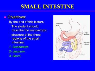
- Objectives
- By the end of this lecture,
- The student should describe the microscopic
- structure of the three regions of the small
intestine - 1- Duodenum.
- 2- Jejunum.
- 3- Ileum.
2
SMALL INTESTINE
- To increase surface area the mucosa has
- Plicae circulares.
- Villi.
- Intestinal crypts (crypts of Lieberkühn).
- Microvilli (Brush border).
3
Duodenum
4
Duodenum
- Mucosa
- Shows villi and crypts.
- A- Epithelium simple columnar epithelium with
goblet cells. - B- Lamina propria C.T.
- C- Muscularis mucosae 2 layers of
smooth muscle cells.
5
Intestinal villi
- Each Villus is a finger-like projection of small
intestinal mucosa and it is formed of - I- Central core of loose C.T. containing
- Lymphocytes.
- Fibroblasts.
- Smooth muscle cells.
- Capillary loops.
- Lacteal (blindly ending lymphatic
channels). - II- Villus-covering epithelium.
6
Cells Covering the Villi
- 1- Surface columnar absorptive cells They have
brush border (microvilli). They are covered wih
thick glycocalx that has digestive enzymes. They
have Junction complex (tight, adhering and
desmosome junctions). - 2- Goblet cells Increase toward the ileum.
- 3- Enteroendocrine cells (DNES cells).
- N.B M cells (microfold cells) They
phagocytose and transport antigens present in the
intestinal lumen. They are mainly found within
epithelium overlying lymphatic nodules of lamina
propria.
7
Intestinal Glands (Crypts)
- Simple tubular glands that open between villi.
- Composed of 5 cell types
- 1. Columnar absorptive cells.
- 2. Goblet cells secrete mucus.
- 3. Paneth cells secrete Lysozymes
(antibacterial). - 4. Enteroendocrine cells secrete hormones.
- 5. Stem cells regenerative cells.
8
Columnar Absorptive cells
Paneth cell
9
Duodenum
- 2. Submucosa
- Connective tissue containing blood vessels
nerves. - Contains Brunners glands (secrete mucus).
- 3. Muscularis Externa
- 2 smooth muscle layers
- Inner circular layer.
- Outer longitudinal layer.
- 4. Serosa or Adventitia
- Except for the 2nd 3rd parts of the duodenum,
which have adventitia, the entire small intestine
is invested by a serosa.
10
Duodenum
11
Regional differences of small intestine
- Duodenum Its submucosa has Brunners glands.
- Jejunum has neither Brunners glands nor
Peyers patches. - Ileum Its lamina propria, opposite the
attachment of the mesentery, has lymphoid nodules
(Peyer's patches) that extend to the submucosa.
12
THANK YOU


