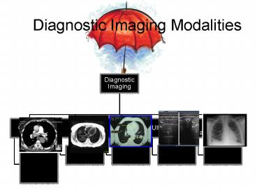
Diagnostic Imaging Modalities - PowerPoint PPT Presentation
Diagnostic Imaging Modalities
The amount of x-ays passing through the hand determine the contrast or how much of the image is black, ... then the risks of contrast nephropathy are negligible. – PowerPoint PPT presentation
Title: Diagnostic Imaging Modalities
1
Diagnostic Imaging Modalities
2
Preparing Images for Viewing
- Images must be labeled for position
- Left /right
- AP/PA/Obl
- Upright, supine, prone
- Patient Data
- Name
- Date
- Account number
3
Viewing an Image
- As though looking at the patient in the
Anatomical Position
4
X-rays pass through a body part and put an image
on a film
5
How is an Image formed?
6
Kubor abd.
right
7
Barium EnemaBE
R
8
Chest PA Lateral
Left
9
CT Scan Transverse Slice of Thorax
10
Elbow anterior posterior position
R
11
Lateral index finger
12
KneeRadiograph (x-ray) MRI
13
Lumbar spineAp Lateral
14
Gastrointestional GI Series
15
ABD.before injectionIVP after
injectionintravenous pyelogram
16
resources
- http//www.netdoctor.co.uk/health_advice/examinati
ons/mriscan.htm - http//en.wikipedia.org/wiki/Magnetic_Resonance_Im
aging - http//en.tnd.kz/equipment/patient_monitoring/obst
etrical_monitoring/ - http//www.radiologyinfo.org/en/photocat/photos_mo
re_pc.cfm?pgbonerad
PowerShow.com is a leading presentation sharing website. It has millions of presentations already uploaded and available with 1,000s more being uploaded by its users every day. Whatever your area of interest, here you’ll be able to find and view presentations you’ll love and possibly download. And, best of all, it is completely free and easy to use.
You might even have a presentation you’d like to share with others. If so, just upload it to PowerShow.com. We’ll convert it to an HTML5 slideshow that includes all the media types you’ve already added: audio, video, music, pictures, animations and transition effects. Then you can share it with your target audience as well as PowerShow.com’s millions of monthly visitors. And, again, it’s all free.
About the Developers
PowerShow.com is brought to you by CrystalGraphics, the award-winning developer and market-leading publisher of rich-media enhancement products for presentations. Our product offerings include millions of PowerPoint templates, diagrams, animated 3D characters and more.


![[PDF] Small Animal Diagnostic Ultrasound Android PowerPoint PPT Presentation](https://s3.amazonaws.com/images.powershow.com/10099022.th0.jpg?_=20240814118)


