SPECT Gantry - PowerPoint PPT Presentation
1 / 18
Title:
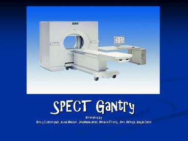
SPECT Gantry
Description:
SPECT Kuhl went on to develop the procedure known as single photon ... the consequences are that leveling the detector head results in data being acquired ... – PowerPoint PPT presentation
Number of Views:221
Avg rating:3.0/5.0
Title: SPECT Gantry
1
- SPECT Gantry
- Presented by
- Besly Clairvoyant, Alina Malave, Jonathan Arias,
Monica Tevez, Joel Ortega, Nayda Carvi
2
History of Nuclear Medicine
- Has its origins in a great many scientific
discoveries dating back to the invention of the
X-ray. - In 1946, radioactive iodine was used to treat a
patients thyroid cancer. - In 1950, radioactive nucleotides were being used
to treat hyperthyroidism. - Soon after, it was possible to record snapshot
images of form and structure or organs ( liver,
spleen, brain, gastrointestinal track, etc.)
3
History of SPECT
- Built his first scanner as a first-year medical
student at Penn. - Edwards and Kuhl developed the MARK VI, the first
Emission Computed Tomography (ECT) device. - MARK VI consisted of several sodium iodide photon
detectors arranged in the rectangular shape
around the head of the patient.
4
SPECT
- Kuhl went on to develop the procedure known as
single photon emission computed tomography
(SPECT) and the principles of positron emission
tomography (PET). - SPECT involves detection of gamma rays emitted
singly from radionuclides such as Tc-99m, I-123,
and I-111. - Tomomatic-32, first SPECT imaging device, was
similar to the MARK VI but has 32 photon
detectors.
5
What is the Gantry?
- The Gantry is the largest part of the SPECT
system. - The Gantry supports the cameras rotation around
the patient, making it possible for images to be
obtain from different angles. - Patients can be easily imaged on hospital beds,
stretchers, or sitting upright, with the help of
the Gantry.
6
Room Requirements
- The room where the camera and Gantry will be
installed must be prepared for this special
equipment. - There must be enough room where the Gantry,
camera, and bed can fit easily within the room
and still allow for other equipment and moving
capabilities. - The floor also has to be able to hold at least
4000 lbs due to the very heavy equipment.
7
Operating the Gantry
- The Gantry can be controlled both by the
computer, and by a hand pendant. - When using the hand pendant the display reads out
the position to aid the operator ( i.e. vertical,
horizontal, or various angles.) - Some Gantry have Intel Pentium based intelligent
Gantry electronics. - The Gantry can be pre-programmed with motions for
positioning.
8
Operating the Gantry
- The Gantry moves along floor rails, using a gear
to both propel itself and calculate its position. - All motions are motorized and computer
controlled.
9
Interesting Fact
- When researching the Gantry, we found another
type of Gantry used in Nuclear Medicine. - In radiation therapy, the Gantry is used for
rotating the radiation delivery apparatus around
the patient, so as to treat from different
angles. - Radiation therapy is the use of high-energy
penetrating rays or subatomic particles to treat
disease.
10
(No Transcript)
11
Gantry Quality Control
- For a tomographic acquisition in single headed
SPECT systems, the gantry is usually set to 0o
and the detector head is leveled prior to an
acquisition. - Setting the detector head level is based on the
assumption that the axis of rotation of the
detector head is horizontal. - The axis of rotation is determined by the
alignment of the gantry.
12
(No Transcript)
13
Quality Control
- Misalignment of the Gantry can be caused by a
number of things. - In many older SPECT systems, sagging of the
detector arms can occur. - The Gantry itself may not be leveled on the
floor. Either due to - Incorrect shimming of the Gantry
- Irregularities in the surface of the floor
14
Quality Control
- Whatever the cause, the consequences are that
leveling the detector head results in data being
acquired obliquely rather than perpendicular to
the axis of rotation. - For example a 1o Gantry misalignment with the
detector head at a 20 cm radius of rotation will
cause a 3.5 mm displacement of image data along
the axis of rotation.
15
Quality Control
- Gantry alignment and its stability with rotation
can also be easily checked using a small bubble
level. - Level the Gantry at 0o, then rotate the Gantry
through 180o and check that the Gantry is still
leveled. - Alignment should be checked once or twice a year
and after any major upgrade or modification to
the Gantry.
16
(No Transcript)
17
Conclusion
- Make sure when setting up a Nuclear Lab that you
have ample room for all equipment. - The SPECT Gantry is a very important part of the
camera. - The Gantry makes it possible to position the
patient for imaging. - It can be operated by computer or by a hand
pendant.
18
- Quality Control is very important on the Gantry
to make sure proper imaging is done. - Make sure to check alignment and leveling.
- Alignment should be checked once or twice a year.
- A Gantry that is misaligned will effect an image,
and Quality Control should be taken very
seriously in regards to the Gantry.

