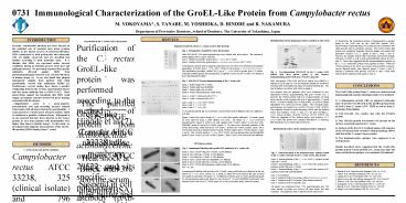
Immunological Characterization of the GroEL-Like Protein from Campylobacter rectus
1 / 1
Title:
Immunological Characterization of the GroEL-Like Protein from Campylobacter rectus
Description:
... from periodontopathogenic bacteria cross-reacted with ... Bacteria were transferred to sterile glass tubes in an anaerobic chamber and sealed with caps. ... –
Number of Views:45
Avg rating:3.0/5.0
Title: Immunological Characterization of the GroEL-Like Protein from Campylobacter rectus
1
- Immunological Characterization of the GroEL-Like
Protein from Campylobacter rectus - M. YOKOYAMA, S. TANABE, M. YOSHIOKA, D. HINODE
and R. NAKAMURADepartment of Preventive
Dentistry, School of Dentistry, The University of
Tokushima, Japan
0731
To characterize the biochemical nature of
immunoreactive epitopes found in the SAM and on
the GroEL-like protein, SDS-PAGE/Western
immunoblotting were performed after treatment
with either periodic acid or proteolytic enzymes.
The 64-kDa GroEL-like protein was completely
degraded when the sample was treated with
proteolytic enzymes (Fig. 5, panels B and C,
lanes 2 and 3). However, the samples treated
with 1 periodic acid reacted strongly with
pAb-CrGroEL as well as with mAb-HuHSP60 (panels B
and C, lane 4). These facts suggested that the
immunodominant epitopes of the C. rectus
GroEL-like protein were peptidic in nature. When
the 150-kDa protein in the SAM was treated with
proteolytic enzymes, several digested bands were
detected (panel A, lanes 2 and 3), whereas a
treatment with periodic acid led to only a faint
band detected at the migration front (panel A,
lane 4).
Recently, considerable attention has been focused
on the potential role of bacterial heat shock
proteins (HSPs) in the disease process of
bacterial infections. HSPs synthesized by both
prokaryotic and eukaryotic cells are highly
conserved, and are classified into families
according to their molecular mass. It is thought
that HSPs are expressed under stressed conditions
during an infection process when host and
pathogen interact. We previously reported that
specific antibodies directed against HSPs from
periodontopathogenic bacteria cross-reacted with
those of human origin (1). It was also found
that gingival homogenate samples from patients
with adult periodontitis reacted with antihuman
HSP60 (2). Furthermore, several studies have
shown a positive relationship between the
severity of periodontal disease and the serum
antibody titer to HSPs (3,4,5). These
observations support the hypothesis that HSPs
could trigger an immune response involved in
tissue damage associated with the disease.
Campylobacter rectus is a gram-negative,
asaccharolytic and agar-corroding bacteria
isolated from lesions with advanced destructive
periodontitis. C. rectus possesses a surface
layer (S-layer) protein, which is considered a
putative virulence factor. Kokeguchi et al. (6)
reported that IgG titers directed to the S-layer
protein in sera from patients with periodontitis
were higher than those in healthy donors. In
this study, we investigated the antigenic
characteristics of the GroEL-like protein (HSP60
family) from C. rectus.
The GroEL-like protein from C. rectus was
characterized immunologically and the following
results were obtained 1) A homogenous protein
with an apparent molecular mass of 64-kDa was
detected in the purified preparations of GroEL
from C. rectus ATCC 33238 as well as from A.
actinomycetemcomitans. 2) PAb-CrGroEL was
reactive also with the 150-kDa protein. 3) This
150-kDa protein was found to be present on the
surface-associated material of C. rectus. 4)
Analysis of the first 20 N-terminal amino acid
sequence of the 150-kDa protein revealed a strong
homology (80) with that of the C. rectus S-layer
protein. 5) The immunoreactive epitopes were
supposed to be carbohydrate. Results described
above suggested that the GroEL-like protein and
the S-layer protein of C. rectus may share the
same carbohydrate epitopes as an antigenic
determinant.
C. rectus strains and growth conditions Campylobac
ter rectus ATCC 33238, 325 (clinical isolate) and
796 (clinical isolate) were grown in trypticase
soy broth supplemented with 0.2 yeast extract,
0.3 phytone peptone, 0.2 NaCl, 0.3 ammonium
formate, 0.4 sodium fumarate and 0.4
L-asparagine adjusted to pH 7.8. Bacterial
cultures were incubated at 37oC in an anaerobic
chamber (85 N2, 10 H2 and 5 CO2) and grown to
mid-log phase (24-36 h incubation). Bacteria
were transferred to sterile glass tubes in an
anaerobic chamber and sealed with caps. Tubes
were removed from anaerobic chamber and
heat-shocked in a water bath at 37oC or 44oC for
6 h. After incubation, C. rectus cells were
chilled in ice, washed with phosphate-buffered
saline (PBS) and placed in 10 trichloroacetic
acid at 4oC overnight. The bacterial cells were
washed twice with PBS and stored at
-20oC. Preparation of the surface-associated
material (SAM) The unstressed cells of C. rectus
ATCC 33238 were washed once with PBS, and then
the SAM was extracted by gentle stirring in
saline for 1h at 4oC. Bacterial cells were
removed by centrifugation at 10,000 g for 20 min.
The supernatant was dialyzed overnight against
distilled water, lyophilized and stored at -20oC.
1. Hinode D et al. Oral Microbiol Immunol 1998
13 55-58. 2. Ando T et al. Microbiol Immunol
1995 39 321-327. 3. Lopatin DE et al. J
Periodontol 1999 70 1185-1193. 4. Schett G et
al. Int Arch Allergy Immunol 1997 114
246-250. 5. Tabeta K et al. Clin Exp Immunol
2000 120 285-293. 6. Kokeguchi S et al. J Clin
Microbiol 1989 27 1210-1217. 7. Hinode D et al.
J Microbiol Methods 1996 25 349-355. 8. Wang B
et al. Infect Immun 1998 66 1521-1526.


![Horizons and Growth Strategies in the Global Campylobacter Testing Market[2016]](https://s3.amazonaws.com/images.powershow.com/8303113.th0.jpg?_=20151118063)


