NanoMEGAS - PowerPoint PPT Presentation
1 / 2
Title:
NanoMEGAS
Description:
... ab ibitio with same resolution like in single crystal X- ray crystallography. ... (Univ of Aachen, Germany) ,Dr X.Zou , S.Hovmoller (Univ of Stockolm, Sweeden) and ... – PowerPoint PPT presentation
Number of Views:42
Avg rating:3.0/5.0
Title: NanoMEGAS
1
NanoMEGAS Advanced Tools for electron diffraction
Blvd Edmond Machtens n79 B-1080 Brussels
Belgium http//www.nanomegas.com
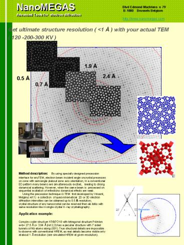
Get ultimate structure resolution ( lt1 Å ) with
your actual TEM ( 120 -200-300 KV )
1.9 Å
2.4 Å
0.5 Å
0.7 Å
a
Method description By using specially designed
precession interface for anyTEM, electron beam
incident angle oncrystal precesses on cone with
semiangle f about zone axis orientation In a
conventional ED pattern many beams are
simultaneously excited, leading to strong
dynamical scattering. However, when the same beam
is precessed on sequential excitation of
reflections dynamical effects are weak.
Using the precession technique in TEM first
developed by Vincent, Midgley( ref.1) a
collection of quasi-kinematical 2D or 3D
electron diffraction intensities can be obtained
up to 0.5 Å resolution crystal structure of any
nanocrystal can be resolved then ab ibitio with
same resolution like in single crystal X- ray
crystallography.
d
Application example Complex oxide structure
KNb7O18 with tetragonal structure P4/mbm ab
27.5 Å c 3.94 Å (ref.2,3) has a peculiar
structure with 7 sided tunnels of Nb atoms along
(001). True structural details are impossible to
observe with conventional HREM, as real details
become visible only at about 1 Å resolution (see
simulated HREM at given resolution).
d
2
NanoMEGAS Advanced Tools for electron diffraction
Blvd Edmond Machtens n79 B-1080 Brussels
Belgium http//www.nanomegas.com
FROM DIFFRACTION PATTERN TO 2D STRUCTURAL
MAP AT ONE STEP
PRECESSION ON
PRECESSION OFF
3600 REFLECTIONS
By using special precession device fitted in a
200KV TEM precession ED pattern resolution
extends dramatically up to 0.5 Å by measuring
precisely quasi-kinematical ED intensities (
electron diffractometry ) and using ab initio
standard direct methods crystallographic
software, complete 2D crystal structure with
all heavy atoms appear in their correct
positions.
PRECESSION TEM interfase SPINNING STAR
With electron diffractometry we can measure,
collect and combine automatically
quasi-kinematical precession electron
diffraction intensities from different zone axis
to one 3D data set , resolving ab-initio 3D
structure from any nanocrystallite
- easily retrofit to any
- TEM 100-300 KV
- precession possible for
- any beam size 300 - 50 nm
References
- precession eliminates false spots due to
dynamical contributions
1.Vincent Midgley Ultramicroscopy 53 (1994)
271 2. Bhinde et al Acta Cryst B 35 (1979) ,
1318-1321 3. Hu et al Ultramicroscopy (1992)
41,387- 397
- software ELD for automatic
- Intensity/symmetry measurement
Acknowledgements We would like to thank for
valuable contibutions to this application note
Dr Thomas Weirich (Univ of Aachen, Germany) ,Dr
X.Zou , S.Hovmoller (Univ of Stockolm, Sweeden)
and Dr Joaquim Portillo (Univ of Barcelona,
Spain).
- automatic 3D structure determination
- with electron diffractometer


