
Microscopic Examination of Stream Biofilms - PowerPoint PPT Presentation
1 / 1
Title:
Microscopic Examination of Stream Biofilms
Description:
... greater magnification. ... the specimen at a higher magnification, rotate the higher power (10x ... viewed at different magnification (note that the eyepiece ... – PowerPoint PPT presentation
Number of Views:31
Avg rating:3.0/5.0
Title: Microscopic Examination of Stream Biofilms
1
Microscopic Examination of Stream Biofilms
This information is intended as a guide for the
examination of the microbial communities within
stream biofilms. For more information about
stream biofilms and to request your free copy of
the poster stream micro-ecology life in a
biofilm, which provides a colourful and richly
detailed view of the microscopic world of stream
biofilms, see www.streambiofilm.org.nz or contact
us on streambiofilm_at_gmail.com
- Collecting Biofilm. Carefully scrape the biofilm
from the surface of a recently collected stream
rock and transfer 5 to 10 ml of stream water into
a small container to form a biofilm slurry. You
can do this in the field .
- Preparation of the microscope. Rotate the
scanning power (x4) objective lens into position,
and using the coarse focus adjustment knob,
position the objective approximately 1 cm from
the lens.
- Prepare the microscope slide. Stir the biofilm
slurry gently and then transfer 3-4 drops of this
solution onto a clean microscope slide. Gently
place a cover slip on top. If you dont have a
cover-slip dont worry! It will still work.
Note Remember to put rocks back as you found
them, as they provide an important habitat for
aquatic organisms
Eyepiece
- Switch on the light source. Then adjust to about
¾ intensity using the light adjuster dial.
- Position the slide. Place the slide into position
on the microscope stage and centre the biofilm
sample under the x4 objective lens using the
stage adjuster.
- Focus on the sample. Bring the slide onto focus
by moving the objective lens away from the slide
using the coarse focus knob. The specimen can
then be brought into sharp focus using the fine
focus knob and the illuminance adjusted with the
iris diaphragm to provide the best view.
Objective lens
Stage
- Try using a greater magnification. To view the
specimen at a higher magnification, rotate the
higher power (10x or 40x) objective lens into the
viewing position while watching from the side to
ensure that the objective does not touch the
slide. The specimen can now be brought into sharp
focus using the fine focus knob. This procedure
can then be repeated to view samples under
progressively higher power lenses.
Iris diaphragm
Light source
On/Off
Tips.
To avoid breaking your slide with the microscope
lens, always focus by moving the lens away from
the slide. When you need to move the lens closer
to the slide do this while watching the
microscope from the side, then look down the
eyepiece and focus while moving the objective
lens away from the slide. A dirty lens will
distort your image. If the lens requires
cleaning, only use special lens paper, or a soft
facial tissue as the lens is relatively soft and
easily scratched. The objective labelled oil
is used to visualise samples under higher
magnification and requires a different approach
to sample preparation, not covered in this guide.
Stage adjuster
Light adjuster
Fine focus knob
Coarse focus knob
Microscopes very widely in their design. This
guide details the recommended procedures for the
use of a standard compound microscope, which in
some cases may be provided on loan (within
Auckland) by contacting g.lear_at_auckland.ac.nz
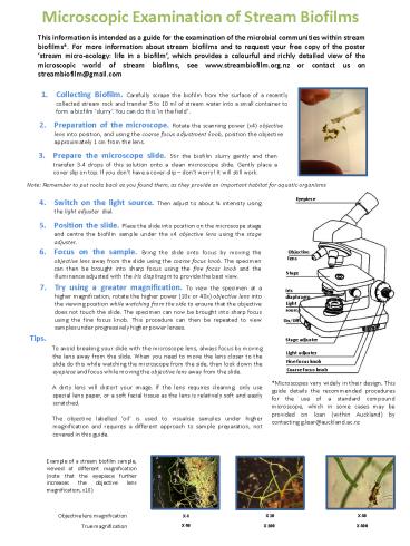
Example of a stream biofilm sample, viewed at
different magnification (note that the eyepiece
further increases the objective lens
magnification, x10)
Objective lens magnification
X 40
X 4
X 10
X 40
True magnification
X 100
X 400
Recommended
CrystalGraphics Presentations


