
Ch 3 first set of notes - PowerPoint PPT Presentation
1 / 14
Title:
Ch 3 first set of notes
Description:
The lighter surround on the left implies more lateral inhibition, resulting in ... Cortical Magnification (packing density) Figure 3.26, page 95 ... – PowerPoint PPT presentation
Number of Views:46
Avg rating:3.0/5.0
Title: Ch 3 first set of notes
1
Ch 3 (first set of notes)
- Psyc 317A
2
Estimating d and ? Corrected Formula
3
Simultaneous contrast
Figure 2.47, page 69
How might apparent darkness difference of the
inner boxes be explained by lateral
inhibition? The lighter surround on the left
implies more lateral inhibition, resulting in
darker appearance. But why dont inner box
centers appear lighter still? Is it legitimate
to sum inhibition over areas?
4
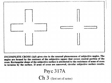
An Aggressive Hermann Gridthe scintillation
effect
5
Pop Quiz
- How many receptor cells are there in each retina?
(approx.) - How many ganglion cells extend axons through/as
the optic nerve? - How many cells are in the Lateral Geniculate
Nucleus? - Where else does the LGN receive input from?
- At what layer of the cortex do LGN neurons
synapse? - How many cells are there in the primary visual
cortex? - What are three other names for the primary visual
cortex? - What is the structure of the retinal fields of
retinal ganglion? - What is the structure of the retinal fields of
LGN neurons? - What is the structure of the retinal fields of
simple cortical cells? - What stimulus features do complex cortical cells
respond best to? - What stimulus features do end-stopped cortical
cells respond best to? - What stimulus variables are represented in a
given hypercolumn of the visual cortex?
6
Lateral Geniculate Nucleus
- The LGN relays the information in exact
point-to-point form - there is a faithful spatial representation of the
on/off pattern of the visual fibers brought from
the retina to the visual cortex - even though the visual tract fibers cross at the
optic chiasm, the LGN is arranged in layers that
keep the signals "parallel" and route the
information from each half of each visual field
to the appropriate cerebral hemisphere. - The LGN also controls how much of the signal
actually gets to the cortex. - Its internal inhibitory circuits can selectively
turn individual signals off and regulate exactly
which visual information is ultimately passed
through to the cortex for processing.
Source Click Here
Note the LGN receives massive input from the
visual cortex, not just from the retina. LGN
neurons also receives input from elsewhere in the
thalamus and LGN
7
Layers and Function in LGN
- nerve cells in layers 1 and 2 are larger than
those in layers 3-6. - Layers 1 and 2 are the Magnocellular layers of
the LGN and receive input from M retinal ganglion
cells - Respond best to movement
- layers 3-6 are the Parvocellular layers of the
LGN and receive input from the P retinal ganglion
cells. - Respond best to texture, colour, pattern, depth,
detail
Each eye provides input to one M layer and 2 P
layers in each LGN
8
Layers and Retinotopic Mapping in LGN
Cells along this line respond to information
coming from the same area of the retina (left or
right) Adjacent receptor fields map to adjacent
neurons in a given LGN layer
- The dark layers on each side contain cells that
respond to stimuli presented to the left eye. - The light layers contain cells that respond to
stimuli presented to the right eye - layers 1, 4, and 6 respond to information from
the contralateral eye, - layers 2, 3, and 5 respond to information from
the ipsilateral eye. SOURCE
9
Columns in Primary Visual Cortex
- Input from LGN arrives in the fourth layer of the
primary visual cortex - There it is processed by simple, complex,
end-stopped and other feature-specific neurons
(e.g. spatial frequency analyzers) - Output goes back to LGN and on to extra-striate
(secondary) visual areas
10
Cortical Magnification (packing density)
- (a) In the fovea, receptors are far more densely
packed than they are in the periphery. - fovea accounts of only 0.01 of retinal area
- (b) Density of ganglion cells is similarly
varied - 50,000/mm2 in fovea
- lt1,000/mm2 off fovea
- (c) Neurons in cortex are evenly spread,
regardless of associated retinal area - fovea maps onto 8-10 of visual cortex
Figure 3.26, page 95
- Therefore, disproportionate share of cortical
processing for vision is dedicated to input from
fovea. - High acuity means high relative intensity of
processing
11
typicalsimple cortical cell receptive field
Figure 3.9, page 83
Neurons in the primary visual cortex also have
receptive fields in the retina. They have both
excitatory inhibitory areas. Cells are usually
orientation specific. Complex cortical cells
often respond best to directional movement across
their field. There are retinotopic maps in both
the LGN and the primary visual cortex.
12
Columns in the cerebral cortex
- Location Columns
- as electrode passes perpendicularly from
surface, the neurons it meets respond to
stimulation from the same general area of of the
retina (over-lapping receptive fields) - if the electrode passes obliquely, it meets
neurons with adjacent (and gradually more
distant) receptive fields - Orientation Columns
- as electrode passes from surface, all the
neurons it meets respond differentially to the
same orientation - Note, as electrode enters perpendicularly from
the cortical surface, it encounters simple,
complex, and end-stopped cortical cells (with the
same orientation tuning and same general
receptive field)
13
Hypercolumns in the visual cortex
- Oracular Dominance Columns
- neurons in the primary visual cortex (V1 or
Brodmann area 17) respond best to stimulus in one
eye - information from corresponding areas of left and
right retinas is processed in nearby parallel
columns - oracular dominance columns are 0.25-0.5 mm wide.
- Orientation Columns
- Within a hypercolumn, there are orientation
specific cells covering all possible orientations
- orientations from all 180 are sampled over ?1
lateral mm
All the cells in a given hypercolumn respond to
stimulation in the same general retinal location
(receptor field)
14
Aspects of the representation of tree in cortex
The representation in neural activity of an
external stimulus is not similar to or like the
object itself. It may not even be
contiguous. Representation does not imply
similarity (cf. diversity in political
representation) Representations of
objects are distributed across neural areas
Figure 3.32, page 99
Recommended
CrystalGraphics Presentations


