Automatic Detection And Classification Of Microcalcifications In Digital Mammograms - PowerPoint PPT Presentation
Title:
Automatic Detection And Classification Of Microcalcifications In Digital Mammograms
Description:
... INFN (National Institute for Nuclear Physics) sections (Bologna, Pisa and Udine) ... has been compiled at Bari and Udine hospitals and contains 2000 images. ... – PowerPoint PPT presentation
Number of Views:73
Avg rating:3.0/5.0
Title: Automatic Detection And Classification Of Microcalcifications In Digital Mammograms
1
Automatic Detection And Classification Of
Microcalcifications In Digital Mammograms
2
Outline of the Project
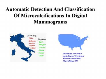
The CALMA Project (Computer Assisted Library for
MAmmography) represents a collaborative effort of
several institutions from Italy and USA INFN
(National Institute for Nuclear Physics) sections
(Bologna, Pisa and Udine), medical centers of
mammographic screening (Bari, Bologna and Udine)
and the Institute for Brain and Neural Systems at
Brown University. Its purpose is to develop a CAD
(Computer Assisted Diagnosis) system for the
detection and classification of mammographic
lesions, based on neural networks and expert
systems. The CAD systems have been quite
successful as diagnosing tools, in many cases
outperforming estimates by expert radiologists.
Our goal is to build a system which consists of a
scanner (or a digital mammogram) and dedicated
hardware and software that can assist
radiologists in their diagnoses. We are currently
developing a software package including tools for
image processing, detection and classification of
lesions. At the moment we are analyzing the most
common lesions in mammograms clustered
microcalcifications. Our data set consists of two
databases. The first database has been compiled
at Bari and Udine hospitals and contains 2000
images. The second database, for evaluation of
our results, is a Nijmegen database which
contains 40 images. Our approach for
preprocessing and image enhancement of digital
mammographs combines classical image and
multiresolution wavelet analysis techniques. Once
the Regions Of Interest (ROI) have been detected,
a set of features characterizing their texture is
extracted from each ROI. Since the number of
texture features is very large, we apply a
feature reduction scheme based on their mutual
correlation and discrimination power. A Neural
Network (NN) based classifier is then trained on
the remaining features in order to separate the
two classes of lesions (benign and malignant
tumors).
3
Possible Types of Lesions
microcalcifications masses
star-shaped lesions (single or
clustered)
4
Microcalcifications
- Microcalcifications look like little spots
(0.1?0.3mm) with high brightness compared to the
surrounding tissue. - They are clinically important if clustered in
groups of three, or more, within an area of about
1cm2.
5
Example of Microcalcifications in a Mammogram


