
AS Unit 2 Energy Transfer Systems Imaging Methods used in Monitoring
1 / 83
Title:
AS Unit 2 Energy Transfer Systems Imaging Methods used in Monitoring
Description:
Radiography & Computed Tomography (CT) Ultrasound (MU / US ) ... a medical imaging technique that uses high frequency sound waves and their echoes. ... –
Number of Views:125
Avg rating:3.0/5.0
Title: AS Unit 2 Energy Transfer Systems Imaging Methods used in Monitoring
1
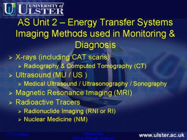
AS Unit 2 Energy Transfer SystemsImaging
Methods used in Monitoring Diagnosis
- X-rays (including CAT scans)
- Radiography Computed Tomography (CT)
- Ultrasound (MU / US )
- Medical Ultrasound / Ultrasonography / Sonography
- Magnetic Resonance Imaging (MRI)
- Radioactive Tracers
- Radionuclide Imaging (RNI or RI)
- Nuclear Medicine (NM)
2
AS Unit 2 Energy Transfer SystemsImaging
Methods used in Monitoring Diagnosis
- Main principles
- Advantages
- Disadvantages
3
Electromagnetic Spectrum
4
(No Transcript)
5
Radiography
6
Stationary Anode X Ray Tube
Heated filament emits electrons by thermionic
emission
Electrons accelerated by a high voltage
Copper for heat dissipation
X-rays emitted when high speed electrons hit the
target
7
Radiography
Rotating Anode x-ray tube
8
Radiography
9
Radiography - Fluoroscopy
10
Radiography
11
Radiography - Fluoroscopy
12
(No Transcript)
13
Radiography
Disadvantages
Advantages
- Ionising radiation
- Does not demonstrate soft tissue well
- Readily available
- Relatively inexpensive
- Relatively quick
- Non-invasive (?)
- Used with/out contrast medium
- Applicable to all parts of skeleton
14
Computed Tomography (CT)
- What is a CT scanner ??
- an x-ray imaging device capable of producing
sectional images - AND
- 3-D reconstructed images
15
Computed Tomography
16
Computed Tomography (CT)
17
(No Transcript)
18
Computed Tomography (CT)
19
Computed Tomography (CT)
20
Computed Tomography (CT)
21
Computed Tomography (CT)
22
Computed Tomography (CT)
23
Computed Tomography (CT)
24
Computed Tomography (CT)
25
Computed Tomography (CT)
Advantages
Small differences in x-ray signals can be
detected Images easily stored retrieved Image
data can be manipulated Relatively
quick Reasonably readily available
Disadvantages
Higher radiation doses than radiography Cost ??
26
Medical Ultrasound (MU)
- Audible frequency range 20Hz 20,000Hz
- Below 20Hz Subsonic
- Above 20,000Hz Ultrasound
27
Medical Ultrasound (MU)
What is Ultrasound - ??
a medical imaging technique that uses high
frequency sound waves and their echoes. The
technique is similar to the echolocation used by
bats, whales and dolphins, as well as SONAR used
by submarines
28
Medical Ultrasound (MU)
Medical Ultrasound (MU)
- The ultrasound machine transmits high-frequency
(1 to 5 megahertz) sound pulses into your body
using a probe. - The sound waves travel into your body and hit a
boundary between tissues - Some of the sound waves get reflected back to the
probe, while some travel on further until they
reach another boundary and get reflected - The reflected waves are picked up by the probe
and relayed to the machine - The machine calculates the distance from the
probe to the tissue or organ using the speed of
sound in tissue and the time of each echo's
return - The machine displays the distances and
intensities of the echoes on the screen, forming
a two dimensional image .
29
Medical Ultrasound (MU)
The Ultrasound Machine
transducer probe central processing unit
(CPU) transducer pulse controls display keyboar
d/cursor disk storage device printer
30
Medical Ultrasound (MU)
Major Uses of Ultrasound
Soft Tissue
the liver the pancreas the gall bladder the
bile drainage system the spleen the kidneys
the tubes running from the kidneys to the
bladder (ureters) the bladder the womb (uterus)
the organs which produce the eggs (ovaries)
Obstetrics and Gynecology Cardiology
Urology Emergency rooms
31
Medical Ultrasound (MU)
3D Ultrasound Imaging
32
Medical Ultrasound (MU)
Doppler Ultrasound - measure the rate of blood
flow through the heart and major arteries
33
Doppler Imaging
34
Medical Ultrasound (MU)
35
MU Imaging
36
Medical Ultrasound (MU)
Dangers of Ultrasound
- some reports of low birthweight babies being born
to mothers who had frequent ultrasound
examinations during pregnancy - development of heat
- bubbles (cavitation)
there have been no substantiated ill-effects of
ultrasound documented in studies in either humans
or animals
37
Magnetic Resonance Imaging (MRI)
38
Magnetic Resonance Imaging (MRI)
Knee MRI - Only modality for picking up meniscal
and cruciate tears Position of Bone bruising in
fat suppressed image is indicative of ACL tear
39
Magnetic Resonance Imaging (MRI)
Brain MRI Can show MS before other modalities due
to ability to saturate fluid from the images and
multi planer images
40
Magnetic Resonance Imaging (MRI)
Magnetic Resonance Angiography Can image the
vessels of the brain without the use of contrast
agent using specialised imaging acquisition like
Time of Flight
41
Magnetic Resonance Imaging (MRI)
Liver (Hepatoma) Has the advantage of being able
to characterise lesions using different pulse
sequences and use of contrast agent
T2
T1 Pre
T1 Post
42
Magnetic Resonance Imaging (MRI)
- Visualising, Evaluating and Diagnosing
- multiple sclerosis (MS)
- tumors of the pituitary gland and brain
- infections in the brain, spine or joints
- tendonitis
- strokes in their earliest stages
- torn ligaments in the wrist, knee and ankle
- shoulder injuries
- masses in the soft tissues of the body
- bone tumors, cysts and bulging or herniated discs
in the spine
43
Magnetic Resonance Imaging (MRI)
Advantages
Disadvantages
- expensive
- too big (patient)
- metal (magnetic)
- pacemakers
- claustrophobia
- noise
- time
- artifacts
- Excellent for soft tissue
- Image reconstruction
- Scan in any plane
- With/out contrast medium
44
Radionuclide Imaging (RNI)
- What is Nuclear Medicine? - the safe use of
radioactive materials in the diagnosis and
treatment of various diseases
- 1. Imaging
- Certain compounds (pharmaceuticals) which
concentrate in different organs of the human
body, are chemically labelled with specific
radioactive materials (radioisotopes) - These radiopharmaceuticals, once administered,
concentrate within the organ or organ system and
the distribution is determined by specialised
equipment.
45
Radionuclide Imaging (RNI)
- 2. Therapy
- certain elements are necessary for the body's
metabolism or normal function - the body cannot distinguish between a radioactive
or non-radioactive element so will deal with both
in the same manner
- 3. Laboratory
- blood products are labelled with specific
radioisotopes and then administered - subsequent sampling of blood or other product or
surface measurements are performed.
46
Radionuclide Imaging (RNI)
Scans performed are many and varied and some
common scans are bone lung renal
(kidney) brain myocardial (heart)
scans.
47
Radionuclide Imaging (RNI)
Radionuclides
Iodine Gallium Thallium Technetium
Technetium has become the most widely used
radionuclide for diagnostic Nuclear Medicine. Its
almost ideal physical characteristics of short
half-life, low energy of its mono-energetic gamma
ray and ease of chelation facilitates its
incorporation into a wide range of
radiopharmaceuticals
48
Radionuclide Imaging (RNI)
Examples of technetium labelled compounds
available and their uses
Radiopharmaceutical Short form Clinical
Use Technetium Sulphur Colloid 99mTcS/CReticulo
Endothelial System (Liver, Spleen and Bone Marrow
Scan) Technetium Macro Aggregated
Albumin 99mTcMAA Pulmonary Blood Flow (Lung
Scan) Technetium Diethylene Triamino Penta
Acetic Acid 99mTcDTPA Renal Blood Flow, Function
and Excretion (Kidney Scan) Technetium
Methylene DiPhosphonate 99mTcMDP Skeletal
Studies (Bone Scan) Sodium Pertechnetate Na299mT
cO4 Thyroid, Salivary Gland and Gastric
Scans 99mTc Red Blood Cells 99mTcRBC Cardiac
Function and Blood Pool Scans 99mTc
Sestamibi 99mTcMIBI Myocardial Perfusion (Heart
Muscle Blood Flow) 99mTc Tetrofosmin 99mTcTETRO M
yocardial Perfusion (Heart Muscle Blood
Flow) 99mTc Hexa Methylene 99mTc HMPAO Brain
Scan and Scans for Infection Propylene Amine
Oxime
49
Radionuclide Imaging (RNI)
The Scan !
- an intravenous injection of a small volume of
radiopharmaceutical - alternative methods of administration are by
inhalation or oral ingestion - depending on the requirements of the scan, the
patient may have images taken immediately or
after a longer period of time, typically 3 - 5
hours - in most cases, all that is required of the
patient is to lie supine (on their back) on the
bed - either the bed will slowly move between the
detectors, the detectors will move or a
combination of both - Gamma Cameras - do not emit ionising radiation -
very sophisticated radiation detectors
50
Radionuclide Imaging (RNI)
51
Radionuclide Imaging (RNI)
Gamma Cameras
52
Radionuclide Imaging (RNI)
Disadvantages
Advantages
Radiation dose
Relatively available Physiology/Function
53
PET-CT
54
Medical Imaging
Further Information
http//science.howstuffworks.com/cat-scan.htm
http//electronics.howstuffworks.com/mri.htm
http//electronics.howstuffworks.com/ultrasound.ht
m
http//science.howstuffworks.com/x-ray.htm
http//science.howstuffworks.com/nuclear-medicine.
htm
55
rheumatoid arthritis
56
rheumatoid arthritis - radiography
57
rheumatoid arthritis - ultrasound
58
rheumatoid arthritis - MRI
59
Conclusion
rheumatoid arthritis - MRI
Radiography is currently the modality of choice
- Is Magnetic Resonance Imaging or Ultrasound the
modality of the future for the detection and
ongoing management for patients with Rheumatoid
Arthritis?
60
Renal Cell Carcinoma (RCC)
Renal Cell Carcinoma (RCC)
- Signs and Symptoms
- Growth and Metastatic Spread
- Subtypes
- Conventional
- Papillary
- Chromophobe
- Collecting Duct
- Unclassified
61
Renal Cell Carcinoma (RCC)
62
Ultrasonography (US)
Renal Cell Carcinoma (RCC)
- Early Diagnosis
- Colour Doppler
- Computed Angiosonography
- Sensitivity
- Limitations
63
Computed Tomography (CT)
Renal Cell Carcinoma (RCC)
- Helical and 3D
- Detection of subtypes
- Secondary
- Sensitivity Specificity
- Surgery
64
Magnetic Resonance Imaging (MRI)
Renal Cell Carcinoma (RCC)
- Alternative to CT
- Advantages
- Sensitivity and Specificity
- Integration with CT US
65
Renal Cell Carcinoma (RCC)
- CT, MU and MRI all have a role to play in the
diagnosis and management of RCC
66
What is Appendicitis?
Appendicitis
- Appendicitis is acute inflammation of the
appendix caused by an obstruction of the
appendix. - Normal Appendix Inflamed Appendix
(www.eMedicine.com)
67
Ultrasound
Appendicitis
- Graded compression ultrasound.
- Primary imaging modality used with paediatrics.
- Fast, reliable, readily available and does not
involve the use of ionising radiation. - Conflicting views on the accuracy of ultrasound.
- Increased sensitivity when used with Doppler.
68
Computed Tomography
Appendicitis
- Use of CT scanning for appendicitis has increased
from 17.6 in 1998 to 51.3 in 2003. - Many advantages over ultrasound.
- Higher reported sensitivity and specificity when
compared with ultrasound. - High radiation dose for paediatrics.
69
Nuclear Medicine
Appendicitis
- Accurate non invasive test for excluding
appendicitis. - Method of labelling WBC with Technetium 99m
Hexamethylpropaline amine oxime - Not suitable for emergency setting.
- Unknown sensitivity and specificity in comparison
with other imaging modalities.
(www.eMedicine.com)
70
Magnetic Resonance Imaging
Appendicitis
- MRI
- Operator Independent
- Non ionising
- Excludes contrast media
- Drawbacks
- Not suitable for emergency setting
- Motion artefact
- Spatial resolution is not comparable to CT or US.
- High cost
- Lack of availability.
www.ajronline.com)
71
Conclusion
Appendicitis
- Medical Imaging
- decrease ve appendectomy rate.
- lower the number of perforations in paediatrics.
- Further research needed for NM and MRI.
- CT vs US
- Specificity and sensitivity higher for CT.
- More pitfalls in US.
- Protocol Combining US
CT
72
BREAST CANCER
BREAST CANCER
- Second leading cause of death in women worldwide
- Most common cancer among females in the UK
(Fig.1) - 12,600 women die in UK each year
- Causes of the disease not fully understood
- No known cure as yet
- Early detection is the key
73
MAMMOGRAPHY
BREAST CANCER
- Currently main detector
- A basic procedure
- NHS carries out screening
- Screening or diagnostic
- Advantage - early identification
- Disadvantage - does not detect all breast cancers
74
THE ROLE OF MRI
BREAST CANCER
- MRI more widely available
- No exposure to radiation
- Provides excellent contrast
- Useful in younger patients
- Not suitable to all patients
- More expensive
75
THE ROLE OF MRI
BREAST CANCER
- MRI has a variety of uses
- More sensitive than mammography
- Contrast enhancement greater visualisation
- Ideal for high risk patients
- Useful after therapy
76
Modality of Choice ?
BREAST CANCER
- Combined use of both modalities
- Better detection rates
- Mortality rates reduced
- Mammography still relevant
- MRI has benefits
- Survival chances are greater
77
Sports Injuries
- Two types of sports injury
- Traumatic
- Chronic
- Incidence of overuse injury in sports has
dramatically risen in past 3 decades. - Increase in participation of sporting events
- Increased intensity of training sessions.
- (Kader et al, 2002)
78
Role of Ultrasound
Sports Injuries
- Ultrasound is an attractive imaging modality for
musculoskeletal imaging due to the widespread
availability, low cost and absence of ionising
radiation. - (Bruce et al, 2003).
- Other modalities can be used
- Magnetic Resonance Imaging
79
Ankle Injuries
Sports Injuries
- Ligament Injury
- Tendon Injury
80
Knee Injuries
Sports Injuries
- Meniscal tears
- Cruciate ligament
- Collateral ligament
- Tendon injury
81
Sports Injuries
- Ultrasound and MRI have critical and pivotal
roles in the management of musculoskeletal
disease. Those who rely on one method and ignore
the other do so at their patients risk. - (Wilson, 2005)
82
Imaging Methods used in Monitoring Diagnosis
- Quite often more than one modality may be used
- Not always a clear favourite
- Choice may be influenced by
- clinical need accuracy, suitability
- availability
- safety
- cost
83
Which do we use?
- What information do we require?
- Do we wish to see function or structure?
- What can the patient tolerate?
- What would the clinician prefer?
- What is available for use?
- Is there a safer/cheaper alternative?
- Can potential risks be justified?
Recommended
CrystalGraphics Presentations


