Chapter 4 Opener - PowerPoint PPT Presentation
1 / 110
Title: Chapter 4 Opener
1
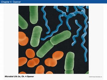
Chapter 4 Opener
2
Figure 4.1 How the human eye magnifies
3
Figure 4.2 Compound light microscope
4
Figure 4.2 Compound light microscope (Part 1)
5
Figure 4.2 Compound light microscope (Part 2)
6
Figure 4.3 Resolving power or resolution
7
Figure 4.3 Resolving power or resolution
8
Figure 4.4 Nonhomogeneous and homogeneous
immersion
9
Figure 4.4 Nonhomogeneous and homogeneous
immersion
10
Figure 4.5 Objective lens
11
Figure 4.6 Dyes
12
Figure 4.7 Various stains used for bacteria and
archaea
13
Figure 4.7 Various stains used for bacteria and
archaea
14
Figure 4.8 Phase contrast microscopy
15
Box 4.1 The Gram Stain
16
Box 4.1 The Gram Stain
17
Figure 4.9 Fluorescence microscopy
18
Box 4.2 The Darkfield Microscope
19
Box 4.2 The Darkfield Microscope (Part 1)
20
Box 4.2 The Darkfield Microscope (Part 2)
21
Figure 4.10 Confocal Microscope
22
Figure 4.10 Confocal Microscope (Part 1)
23
Figure 4.10 Confocal Microscope (Part 2)
24
Figure 4.11 Confocal laser microscopy
25
Figure 4.12 Natural community
26
Figure 4.13 Transmission electron microscope
(TEM)
27
Figure 4.14 Illumination in light and electron
microscopes
28
Figure 4.14 Illumination in light and electron
microscopes (Part 1)
29
Figure 4.14 Illumination in light and electron
microscopes (Part 2)
30
Figure 4.14 Illumination in light and electron
microscopes (Part 3)
31
Figure 4.15 Resolution in light and electron
microscopy
32
Figure 4.15 Resolution in light and electron
microscopy
33
Figure 4.16 Thin section of a gram-negative
bacterium
34
Figure 4.17 Scanning electron microscope (SEM)
35
Figure 4.18 SEM-elemental analysis of manganese
and iron
36
Figure 4.18 SEM-elemental analysis of manganese
and iron
37
Figure 4.19 Atomic force microscope
38
Figure 4.20 Atomic force microscopy
39
Figure 4.21 Typical bacterial and archaeal shapes
40
Figure 4.22 Coccus and rod shape showing binary
transverse fission
41
Figure 4.23 Bacilli, or rods
42
Figure 4.24 Curved and helical cells
43
Figure 4.25 Prosthecate bacterium
44
Figure 4.26 Mycelial bacterium
45
Figure 4.27 Filamentous bacteria
46
Figure 4.28 Cross section of bacterial cell
envelopes
47
Figure 4.28 Cross section of bacterial cell
envelopes (Part 1)
48
Figure 4.28 Cross section of bacterial cell
envelopes (Part 2)
49
Figure 4.29 Appearance of DNA by electron
microscopy
50
Figure 4.30 DNA strands released from cell
51
Figure 4.31 Supercoiled DNA
52
Figure 4.32 Protein synthesis
53
Figure 4.32 Protein synthesis
54
Figure 4.33 Ribosome structure
55
Figure 4.33 Ribosome structure (Part 1)
56
Figure 4.33 Ribosome structure (Part 2)
57
Figure 4.34 Dipicolinate
58
Figure 4.35 Sporulation of an endospore-forming
bacterium
59
Figure 4.35 Sporulation of an endospore-forming
bacterium
60
Figure 4.36 Sporulation process
61
Figure 4.36 Sporulation process (Part 1)
62
Figure 4.36 Sporulation process (Part 2)
63
Figure 4.37 Clostridium tetani
64
Figure 4.38 Gas vacuoles and gas vesicles
65
Figure 4.39 Gas vesicles
66
Box 4.3 The Hammer, Cork, and Bottle Experiment
67
Figure 4.40 PHB granules
68
Figure 4.41 Polyphosphate granules
69
Figure 4.42 Sulfur granules
70
Figure 4.43 Cell diagram
71
Figure 4.44 Bacterial cell membrane structure
72
Figure 4.44 Bacterial cell membrane structure
73
Figure 4.45 Phospholipid
74
Figure 4.45 Phospholipid
75
Figure 4.46 Sterols and hopanoids
76
Figure 4.47 Archaeal cell membrane structure
77
Figure 4.47 Archaeal cell membrane structure
(Part 1)
78
Figure 4.47 Archaeal cell membrane structure
(Part 2)
79
Figure 4.48 Cell walls of gram-positive and
gram-negative bacteria
80
Figure 4.49 Peptidoglycan of a gram-positive
bacterium
81
Figure 4.49 Peptidoglycan of a gram-positive
bacterium
82
Figure 4.50 Diamino acids
83
Figure 4.51 Teichoic acids
84
Figure 4.51 Teichoic acids (Part 1)
85
Figure 4.51 Teichoic acids (Part 2)
86
Figure 4.52 Teichuronic acids
87
Figure 4.53 Pseudomurein
88
Figure 4.54 Protoplasts
89
Figure 4.55 Outer membrane
90
Figure 4.56 Polysaccharide portion of LPS
91
Figure 4.57 Lipoprotein structure
92
Figure 4.57 Lipoprotein structure
93
Figure 4.58 Porin
94
Figure 4.59 Capsule stain
95
Figure 4.60 Negative stain
96
Figure 4.61 Gliding motility
97
Figure 4.62 Sheathed bacteria
98
Figure 4.63 S-layers
99
Figure 4.64 Polar flagellum (monotrichous
flagellation)
100
Figure 4.65 Polar flagellar tufts (lophotrichous
flagellation)
101
Figure 4.66 Peritrichous flagellation
102
Figure 4.67 Flagellar structure
103
Figure 4.67 Flagellar structure (Part 1)
104
Figure 4.67 Flagellar structure (Part 2)
105
Figure 4.67 Flagellar structure (Part 3)
106
Figure 4.68 Phototaxis
107
Figure 4.69 Fimbriae
108
Figure 4.70 Pili
109
(No Transcript)
110
(No Transcript)

