Imaging the Structure of Mouse Embryonic Stem Cells
1 / 1
Title: Imaging the Structure of Mouse Embryonic Stem Cells
1
Imaging the Structure of Mouse Embryonic Stem
Cells
Judith A. Newmark, Robert J. Crooker, and Carol
M. Warner
Bernard M. Gordon Center for Subsurface Sensing
and Imaging Systems Department of Biology
Northeastern University, Boston, MA 02115
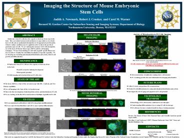
Activity of Mitochondria in ES Cells Imaging
MULTICOLOR IMAGING
ABSTRACT
Embryonic stem (ES) cells have the capacity
to form any tissue type in the body. They are
isolated and grown from blastocyst stage embryos
on a feeder layer of cells and are maintained
through the use of appropriate growth media.
Selective culture conditions may be applied in
order to direct the growth of particular types of
cells. We are studying the structure of ES cells
through the use of the Keck 3D Fusion Microscope
(3DFM) and live cell imaging techniques. We have
been labeling ES cells with organelle-specific
fluorescent dyes in order to visualize the
subcellular localization of specific organelles.
We are focusing on correlating mitochondrial
distribution and activity with ES cell growth and
differentiation.
Multicolor Fibroblasts
Multicolor ES Cells
SIGNIFICANCE
Bluenuclei, GreenJC-1 green (inactive
mitochondria), RedJC-1 red (active mitochondria)
- Embryonic Stem (ES) Cells have the capacity to
form any tissue in the body - model system for studying development
- therapeutic potential
- Little imaging and structural data on ES cells
exists
ES cells contain active and inactive
mitochondria in different locations and with
different morphologies
CONCLUSIONS
- 3D reconstructions of multicolor staining shows
cell structure - JC-1 staining provides the most information for
imaging mitochondria
STATE OF THE ART
FUTURE PLANS
- The Keck 3DFM is a State-of-the-Art microscope
with DIC, Confocal, and Two-Photon capabilities - Live-cell imaging is the State-of-the-Art in
microscopy - This is the first investigation of mitochondrial
activity and distribution in ES cells - We are working on the first 3D reconstructions
of multiple organelles in ES cells
- Collect Z stacks and perform 3D reconstructions
- Predict ES cell differentiation by using
mitochondrial distribution and activity - Investigate the role of aging and apoptosis in
ES cell potential
DIC, Hoechst (nuclei) Blue, JC-1 (inactive
mitochondria) Green, JC-1 (active mitochondria)
Red, Overlay
MITOCHONDRIAL IMAGING
TECHNOLOGY TRANSFER
ES CELLS
DIC
Hoechst (nuclei)
MitoTracker Green
Fluorescence Overlay
- Knowledge of ES cell structure could lead to new
therapies - Understanding differentiation is key to using ES
cells for therapy - ES cells may become a standard model in drug
screening programs
- ES are maintained on fibroblasts with LIF to
keep them undifferentiated - ES are removed from fibroblasts and LIF to cause
random differentiation
REFERENCES
Review The Future of Stem Cells. Financial Times
and Scientific American special report
(2005) Kiessling AA and Anderson S (2007) Human
Embryonic Stem Cells. Jones and Bartlett
Publishers, Inc.
CONTACTS
Judith Newmark (617) 373-3973 j.newmark_at_neu.edu
Biology Dr. Carol Warner (617)
373-4036 c.warner_at_neu.edu Biology
Figure DIC images of ES cells were taken at all
available magnifications on the Keck 3DFM. The
4x and 10x objectives were adjusted to collect
pseudo-DIC images as the objectives are not
capable of performing true DIC.
This work was supported in part by CenSSIS, the
Bernard M. Gordon Center for Subsurface Sensing
and Imaging Systems, under the Engineering
Research Centers Program of the National Science
Foundation (Award Number EEC-9986821) and the
W.M. Keck Foundation.


