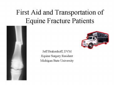
First Aid and Transportation of Equine Fracture Patients - PowerPoint PPT Presentation
1 / 40
Title:
First Aid and Transportation of Equine Fracture Patients
Description:
adobe-xap-filters esc='CR' ... xmlns:xapMM='http://ns.adobe.com/xap/1.0/mm/' xapMM:DocumentID adobe:docid:photoshop:76c4ea12-8e97-11da-95ff-837db5f2df6 6 ... – PowerPoint PPT presentation
Number of Views:434
Avg rating:3.0/5.0
Title: First Aid and Transportation of Equine Fracture Patients
1
First Aid and Transportation of Equine Fracture
Patients
- Jeff Brakenhoff, DVM
- Equine Surgery Resident
- Michigan State University
2
Emergency Management
- Tremendous advancement in fracture fixation
- Successful outcome
3
Emergency Management
- Expensive endeavor
- Proper temporary stabilization influences the
prognosis significantly
4
Prognosis
- Location The above the carpus-tarsus rule
- in adult horses
- Severity
- Age/Weight
5
Prognosis
- Open/Closed
- Configuration
- Soft tissue damage
6
Prognosis
- Intended use
- Temperament
Performance
Breeding
7
- Most of these injuries cannot be managed easily
in the field - Necessitates transport to a surgical facility
8
Emergency Steps
- Calm the patient
- Perform a cursory exam
- Apply protective splint, bandage or cast
- Perform other diagnostics
- Begin therapy
- Transport the patient to a hospital
9
Calm the patient
- Do not move horse
- Lip twitch
- Tranquilization, Sedation, and Pain Relief
- Detomidine/Butorphanol
- Xylazine/Butorphanol
- Goal Relaxation without ataxia
10
Cursory Exam
- Physical exam
- Most are painful and anxious
- Sweating and shocky
- Hemorrhage rarely a problem
- Assess the extent of damage
- Some limbs are too severely damaged for an
attempt at repair
11
Decision
- Discuss findings and prognosis with owner
- Use teaching hospitals as a resource
- Repair vs. Euthanasia
12
Apply Protective Bandage, Splints, and/or Cast
- Objectives
- General guidelines
- Specific fractures and proper application of
splints, etc
13
Objectives
- Preserve the neural and vascular element of the
limb - Prevent an open fracture
14
Objectives
- Protecting the limb from contamination
- existing skin opening
- Stabilization of the limb
- to relieve the anxiety
- Minimize further damage
- bone ends and soft tissue
15
Guidelines
- Bandage
- Apply counter pressure to the injury site
- Swelling
- Protects the soft tissues
- Splints
16
Guidelines
- Splints
- Lightweight
- Durable
- Easy to apply
- Fits snugly
- Prevents damage to soft tissues
17
Splints
- Examples
- Wood
- PVC
- Rebar
18
Splinting
- Techniques vary with fracture location
- Four Zones
19
Zone 1
- Phalanges and distal 1/3 cannon bone
- Dominated biomechanically by the angle of the
fetlock - Fracture site becomes principle bending focus
- Goal Counteract this bending force
20
Phalanges and Distal Cannon Bone
- Align phalanges in same plane as cannon bone
21
Zone 1
Dorsal Splint -with non-elastic tape
Light Bandage
22
Cortical alignment
- Leg Saver
- Kimzey Splint
23
Zone 2
- Distal radius, carpus to midcannon
- Robert-Jones bandage is essential
24
Robert-Jones Bandage
- Applied in multiple layers
- Roll cotton
- Elastic gauze
- Each layer no greater than 1 inch
- Prevents shifting and compacting
- Evenly distributed over limb
- Goal 3x the diameter of the limb
25
Zone 2 -forelimb
- Apply splint over bandage
- Non-elastic adhesive tape
- Caudally and laterally at 90
- Extend from elbow to ground
26
Zone 2 hindlimb
- Similar principles as forelimb
- Less extensive Robert-Jones bandage applied
- Splints
- Caudally and laterally at 90
27
Zone 3 -Radius
- Difficult to stabilize
- Principle musculature lies on lateral side
- Thin skin medially
- Muscles act as abductors
- Preventing abduction is essential
28
Radius
- Robert-Jones Bandage
- Lateral splint
- Extending proximally toward withers
- Fit snug to triceps
- Taped securely at level of axilla
29
Zone 3 -Tibia and Tarsus
- Again difficult to stabilize
- Similar musculature location as forelimb
- Soft tissue damage a concern medially
30
Tibia and Tarsus
- Goal prevent abduction and over-riding
- Robert-Jones bandage up to stifle
- Thick and tight
- Lateral splint
- Bent to follow the contour of limb
- Extend from ground to rump
- Cranial/Caudal splints
- Difficult to apply and of little support
31
Zone 4 -Elbow
- Unable to extend limb effectively
- Loss of triceps apparatus
- Exception to carpus-tarsus rule
32
Elbow
- Goal
- Keep carpus in extension
- Use limb for support and balance
- Full limb bandage
- Caudally applied splint
- Ground to point of elbow
33
Zone 4 Humerus/Scapula
- Well protected by muscle
- Swelling gives some support
- Additional bandaging difficult and not needed
- concerned about radial nerve damage
34
Zone 4 Femur/Pelvis
- Well protected
- Difficult to stabilize
- Support the opposite limb
35
Zone 4 Femur Foals / Minis
Carpus-tarsus rule does not apply to foals
36
Other Diagnostics/Treatment
- Radiographs
- Antibiotics
- Analgesia
- IV fluids
37
Transportation
- Support is essential
- Direction of horse
- Forelimb fracture face back
- Hindlimb fracture face forward
38
Summary
- Location
- Configuration
Comminuted
Soft tissue damage
Open Tibial Fracture
Adult Radial and Femur Fracture
39
Summary
- Age/Weight
- Proper bandaging
- Apply splints at 90
- Support while shipping
40
Thank You
- Fort Dodge
- MSU Surgeons
- Michigan Veterinary Conference


