Quantification of Circulating Cell free DNARNA - PowerPoint PPT Presentation
1 / 14
Title:
Quantification of Circulating Cell free DNARNA
Description:
Mitochondrial DNA copy number was shown to increase in several types of cancer ... Next, cell free genomic and mitochondrial DNA and RNA plasma levels were ... – PowerPoint PPT presentation
Number of Views:234
Avg rating:3.0/5.0
Title: Quantification of Circulating Cell free DNARNA
1
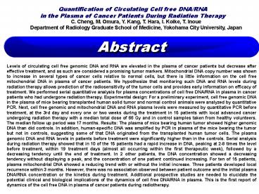
Quantification of Circulating Cell free DNA/RNA
in the Plasma of Cancer Patients During
Radiation Therapy
C. Cheng, M. Omura, Y. Kang, T. Hara, I. Koike,
T. Inoue Department of Radiology Graduate School
of Medicine, Yokohama City University, Japan
2
(No Transcript)
3
(No Transcript)
4
(No Transcript)
5
Table 1, Human specific DNA in the plasma sample
of mice bearing human tumor
Fig. 1-a. Eight of 16 plasma samples of mice
bearing human tumor contain human special genomic
DNA (average 133.3 228.7 ng/mL). None of 10
samples of control mice contains human specific
DNA.
6
To clarify the originated source of cell free DNA
in plasma, mouse specific DNA in plasma samples
were also measured both in mice bearing human
tumor (n16) and control mice (n10). As shown in
Fig.1-b, plasma samples of mice bearing human
tumor showed higher levels of mouse specific DNA
concentration, compared with control mice group.
This result suggests that disintegration of
non-tumor cell in human tumor tissue associated
with tumor growth. It is also possible that
normal tissue around tumor may be destroyed by
tumor invasion.
Fig. 1-b. Plasma samples of mice bearing human
tumor contain more amount of mouse DNA than that
of control mice (average 59.89 64.46 ?g/mL vs
6.72 7.02 ?g/mL). The difference is
statistically significant (plt0.05).
7
Next, human and mouse specific DNA in plasma
sample were compared in mice bearing human tumor.
As shown in Fig.1-c, there was a close
correlation between the concentrations of human
and mouse DNA when the samples contained
detectable human specific DNA (closed circle, ?).
It is suggested that both types of DNA from tumor
and non-tumor tissue were released into the
plasma.
Fig. 1-c The lines show the close correlation
between the concentrations of human and mouse
specific DNA in the plasma of mice bearing human
tumor. (?, the samples contained detectable human
specific DNA, ?, the samples which did not
contain human DNA) Red line was calculated from
all samples (? and ?), R2 0.6136. Green line
was from only samples contained human specific
DNA (?), R2 0.8225.
Cell free mice and human specific mRNA in plasma
samples of mice bearing human tumor were also
analyzed by RT-PCR. However, in those samples,
amplification could not be detectable in our
experimental condition.
8
2. The difference of cell free nucleic acid in
plasma of cancer patients and normal control
persons.
Quantitations of 100 bp or 400 bp genomic DNA,
mitochondria DNA, and mRNA in plasma were
processed by PCR and RT-PCR, respectively. The
DNA concentrations of plasma samples of 15 cancer
patients before radiation therapy were compared
with those of 20 normal control persons.
Patients characters were shown in Table 2. As
shown in Figs.2-a, b, d, e, concentrations of 100
bp or 400 bp DNA, mitochondria DNA and mRNA in
patients samples were higher than those of
normal control persons. The differences were
statistically significant except for 400 bp DNA
(Fig.2-b). Those data suggests the validity of
those types of DNA and mRNA in plasma as a
potential diagnostic marker of cancer.
Fig2-a Concentration of 100 bp genomic DNA in
plasma samples of cancer patients (n15) are
higher than those of normal control persons
(n20) (average 5183.3 8762.6 ng/mL vs
1136.6 1334.6 ng/mL). The difference is
statistically significant, plt0.05).
Fig2-b Concentration of 400 bp genomic DNA in
plasma samples of cancer patients (n15) are
higher than those of normal control persons
(n12) (average 3482.1 5464.1 ng/mL vs 503.8
817.1 ng/mL).
9
The fraction of cell free genomic 100 bp and 400
bp DNA were compared between the samples of
cancer patients and normal control persons. As
shown in Fig2-c, ratio of 400 bp DNA to 100 bp
DNA was increased in plasma samples of cancer
patients compared with those of normal control
persons. This result suggests that DNA integrity
in plasma of cancer patients is increased in
comparison of normal control persons.
Fig2-c The relationships between 100 bp and 400
bp genomic DNA in plasma of cancer patients (?)
and normal control persons (?). Ratio of 400 bp
DNA to 100 bp DNA was increased in plasma samples
of cancer patients compared with those of normal
control persons.
10
Fig2-d Concentration of mitochondria DNA in
plasma samples of cancer patients (n15) are
higher than those of normal control persons
(n20) (average 257.2 435.2 ?g/mL vs 38.9
46.2 ?g/mL). The difference is statistically
significant (plt0.05).
Fig2-e Messenger RNA concentration levels in
plasma of 15 cancer patients are higher than
those of 19 normal control persons (average
645.9 967.4 vs 97.2 137.4). The difference is
statistically significant (plt0.05). cDNA
reverse-transcribed from 2 ?g mRNA of exponential
growing HeLa cell was used as standard of 10000.
11
(No Transcript)
12
Three of 15 patients developed local recurrence
disease and four patients did distant metastasis.
However, there was no association observed
between patient outcome and the initial plasma
DNA and mRNA concentrations or the kinetics
during treatment.
Table 2, Characters and Variation patterns of
DNA concentration during radiation therapy in 15
cancer patients
13
(No Transcript)
14
Fig. 3 Variations in plasma 100 bp, 400 bp, and
mitochondria DNA concentration in cancer patients
treated with radiation therapy and sampled weekly
during the course of treatment (100 bp, top 400
bp middle mitochondria bottom panels). The scale
of Y axis is the plasma mitochondria DNA levels
in percentage, and the X axis is days after the
start of radiation.


