Apoptosis - PowerPoint PPT Presentation
Title: Apoptosis
1
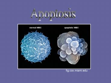
Apoptosis
fig.cox.miami.edu
2
Background
- Cell suicide
- Construction,
maintenance,
repair - All nucleated
cells
ghr.nlm.nih.gov
3
- Vogt 1842
- J.F. Kerr et al (1972)
- Genetically controlled
- Molecular activators
- Evolutionarily conserved
C. Elegans expasy.org
4
- 4 functional group
genes - Ced-3 ? caspases
- Ced-4 ? Apaf-1
- Ced-9 ? Bcl-2
- Egl-1 ? BH3
5
- Human viral, degenerative diseases
- Therapy - cancer
2006 Wikipedia CD Selection
6
Embryonic development
- Physiologically, genetically controlled
- Malformations
- Different stages, organs
- Sculpts organs
morphogenetic - Removal of cells -
histogenetic
www.ccs.k12.in.us
7
Appendages
- Mesoderm
- Amount of cells for
skeleton - Between digits
- Chondrogenetic skeletal
condensations
8
- Ectoderm
- Apical ectodermal
ridge (AER) - Mesenchymal cells
www.tmd.ac.jp
9
Fish
- Many organs
- Sensory organs, brain morphogenetic
- Fins epidermal cells, cartilage
- Median fin fold unpaired dorsal, anal, caudal
10
- 20 hrs post fertilization (hpf)
median fin fold appears - 22 hpf distal parts of fin
fold, proximally - 24 hpf distal tip
- Present until 72 hpf
- Not morphogenetic
Hpf Median fin fold
12 14 16 - - -
20 22 24 0 11.0 /- 5.57 7.00 /- 6.08
30 36 48 13.67 /- 13.50 20.00 /- 6.24 12.00 /- 6.00
60 72 20.33 /- 8.08 5.00 /- 2.83
Apoptosis in median fin fold Cole and Ross, 2001
11
Amphibians
- Not with free digits
- Some anurans, urodeles
absent - Some salamanders
similar to amniotes - X. laevis different proliferation rates in
digital, interdigital - Hindlimb comparable
12
Reptiles
- Patterns correlate with adult
limb morphology - Turtles distal interdigital
areas - Lizards interdigital
- Apoptosis digit formation
1st in amniotes (Fallon and
Cameron, 1977)
13
- Ectoderm of AER
- Cells undifferentiated, proliferating
- Snakes massive apoptosis
14
- Chameleon
- Autopodial cleft
- Specialized
interdigital cell death - Begins early, wide
along distal margin
15
Avians
- Skeletal
primordia of
limbs - 2 areas undifferentiated mesenchyme
- Anterior, posterior margins proximal segment of
limb - Anterior, posterior necrotic zones (ANZ, PNZ)
- Reduction in digit number
www.dls.ym.edu.tw
16
- Opaque patch (OP) central mesenchyme
- 2 pieces of zeugopod
- Digit formation -
mesenchyme
between rays - Interdigital necrotic
zone (INZ)
www.dls.ym.edu.tw
17
- Constant w/in species
- Different for different
species - Correlate w/ limb
morphology - Free digits throughout interdigital space
- Webbed feet distal interdigital
- Free digits
membranous fold
central interdigital
18
- Inhibited syndactyly
- AER spatial, temporal extension of limb
Kingfisher www.turtletrack.org
19
Mammals
- Ectodermal
morphogenetic - AER postaxial, preaxial
margins - Regression of extreme ends
- Inhibition - polydactyly
www.nature.com
20
- Postaxial ridge digit V
- Interdigital digit IV
- Later, entire length
- Decreases except digit I
www.gsc.riken.go.jp
21
- Footplate foyer preaxial primaire (fpp)
- Similar to ANZ
- Reduces quantity of
preaxial mesodermal cells - Talpa (mole) fpp absent,
falciform digit
fpp
www.nature.com
www.palaeos.com
22
- Subridge mesoderm foyer marginal I (fmI)
preaxial margin - Digits 1-3 in forelimb
- 1-1/2 digit 2 in hindlimb
- Foyer marginal V (fmV) postaxial
margin - Digit 5 to border of 4
- Growth of digital buds
- Decrease in influence of ectodermal layer
fmI
fmV
23
- Interdigital mesoderm
separation of digits - 2 waves superficial layer of subectodermal
cells - Between precartilaginous
rudiments of phalanges
24
Mechanisms
- Bone morphogenic proteins (Bmps)
- Transforming growth factor ß superfamily
- Bmp-2, 4, 7, 5 undifferentiated limb mesoderm,
interdigital mesoderm, AER - Coincide with cell death
25
- High redundancy
- Regulated by Bmp
antagonists - Noggin, gremlin,
DAN, Drm - Gremlin ducks, down regulated in chicks prior
to INZ
26
- Bmps limb patterning,
regulate chondrogenic
differentiation - Signal
serine/threonine
receptor kinase with
type I and II receptors - Binding association of 2 receptors
- Phosphorylation of type I by II
- Propagation of intracellular signal
www.medscape.com
27
- Chondrogenic effects
type Ib receptor - Type Ia control of
apoptosis - Interdigital induction
of Ib ectopic digit
arthritis-research.com
28
- Bmps signal through
Smads - Bmp binds to receptor
- Smad cascade
BMP-responding
smads 1, 5, 8 - Co-smad 4
- Inhibitory smads 6, 7
- Translocated to nucleus
- Activate transcription
29
- Also signal through
MapK pathway - Erk, Jnk, p38 kinase
mediate Bmp
signaling
MapK pathways e-kisstoth.staff.shef.ac.uk
30
- Limb
caspase-3, 9, 2 - Death
Inducer-Obliterator-1
(DIO-1) - Growth Arrest
Specific1, 2 (Gas1, 2) - Apaf-1
arthritis-research.com
31
- Bax Bcl-2 family
proapoptotic - Antiapoptotic Bcl-2,
Bcl-x, A1 digital rays
not interdigits - Before apoptosis Bag-1 expresses antiapoptotic
protein - Binds to Bcl-2 in interdigits
- Defender Against apoptotic cell Death (Dad-1)
- Syndactyly
32
- Fgf signaling
outgrowth of limbs - Cooperate with
Bmps - Blocked Bmps do
not trigger
apoptosis - Webbed feet of
ducks decrease
in Fgf - Fgfs activate ERK
www.nature.com
33
- Retinoic acid signaling
limb patterning - Acts with Bmps
interdigital regions - Promotes apoptotic
effects of Bmps - Inhibits chondrogenic
effects - Bmps induce
apoptosis, promote
cartilage growth
a-b dying cells c-d macrophage distribution e-f
macrophage specific antibodies g-h S phase
nucei Dupe et al, 1999
34
Joint Formation
35
- Internal skeleton support, locomotion
- Joints classified by structure, degree of
movement - 1. synarthrosis joined by cartilage
- 2. schizarthrosis interzone contains single
(small ) of cavities - 3. hemiarthrosis single joint cavity, elements
united around perifery - 4. eudiarthrosis separate
articulating elements, cavity limited by
synovial tissue
36
- Degree of movement a joint
allows - 1. Synarthrosis
no movement - 2. Amphiarthrosis limited
movement - 3. Diarthrosis freely movable
37
- Diarthroidal joints aquatic to terrestrial life
- Agnatha to
Gnathostomata
hinged mandible - Greater range of prey
38
- Agnathans branchial
arches - Mandibular arch
chondrocranium
jaws - Upper palatoquadrate
lower mandibular cartilage - Mammals - malleus incus
- diartroidial
39
- Chondrichthyes
- Charchariniformes,
Squaliformes
hemiarthrosis - Holocephali more
diarthroidial - Synovial membrane on
one side - More analysis
40
- Rajidae diarthroidial
- Arose in cartilaginous fish
- May be lost in
elasmobranches
41
- Osteichthyes some
may have lost diarthroses - Quadrate/mandibular -
microscopic structure - Polypterus, Protopterus
Haines, 1937
42
- Lepidosteus (longnose
gar) - Layer of calcified cartilage
- Hypertrophic chondrocytes
integrated into bone - Hyaline cartilage
- Articular fibrocartilage birds, mammals
- No fibrous capsule loose
connective tissue - Synovial membrane
bilayered
AF
CC
HC
Haines, 1942
43
- Early dipnoans bony, supported by overlying
cartilage - Living secondary modification
- Fins - no diarthrosis
- Synarthrosis distal, smaller
joints - Schizoarthrosis, hemiarthrosis
proximal, larger joints
44
- Amia calva (bowfin)
proximal radial/girdle
diarthroidal? - Joint cavity, minimal
connective tissue,
2-layered synovium - Modern bony fish more diarthroses
- Maneuverability swimming, feeding
- Larger size large joints at base of fins
45
- Tetrapod limbs
diarthroidial - Urodeles, anura
distal joints
synarthroses - May be secondary modification
46
- Primitive structure
no joint capsule
supporting 2-layered
synovial membrane - Amphibia, reptilia
- Fibrous/fibrocartilaginous
articulating surface overlying hyaline cartilage
of epiphysis - birds
Crocodile knee Haines, 1942
47
- Crocodilus, Sphenodon,
lizards primitive - Cruciate ligaments,
menisci, single joint
cavity femur/tibia/fibula - Chelonians firm
articulation between
median condyle of
femur/tibia - Reduction in medial meniscus
48
- Urodeles reduced/lost cavity, menisci,
ligaments - Marsupials,monotremes
femora-fibular
articulation - Joint cavity subdivided
by connective tissue - Eutherians articulation
lost - Femur closely bound to
tibia
49
- Histological features of joint epiphyses
- Bony fish
cartilaginous
epiphyses at end
of diaphyses - Articular surfaces fibrous
- Perichondrium?
- Mass of rounded chondrocytes surrounded by
cartilage matrix - Metaphyses become flattened, hypertrophied
50
- Matrix may become
calcified - Reabsorbed by elements
of bone marrow marrow
processes - True endochondral
ossification
51
- Calcified cartilage in
center of epiphyses of
epibranchial bone - Forerunner of secondary
center of ossification - Closing plate of
endochondral bone - Bony fish true epiphysis endochondral growth
mechanism
52
- Early tetrapods
cartilaginous epiphyses - Lacked secondary centers
of ossification - Chelonia, Crocodilia retain
primitive condition - Modifications for land dwelling
- Reduction in zone of round cells
- Flattened zone closer to articular surface
- Firmer epiphysis
53
- Tuatara most primitive
secondary ossification - Large masses of calcified
cartilage - Greater part of adult
epiphysis - Thin layer of articular cartilage
- Flattened cell zone partitioned into columns
- Division of founder population at top of column
- Progeny lie beneath mother cell
54
- Mammals alignment
occurs early - Reptiles/birds initially
not aligned - Loose alignment in
postembryonic development - Maintained during hypertrophy, calcification
- Only septa between hypertrophied cells calcifies
- Form templates for endochondral bone
55
- Noncalcified septa
broken down by
metalloproteinases - Hypertrophs undergo
apoptosis,
transdifferentiation into
ostoblasts
56
- Cavity greater range of
motion - Schizarthrosis primitive
condition? - Mechanism differential
hyaluronan (HA) synthesis - Mechanical stimuli
- Glycosaminoglycan HA, CD44 differentially
expressed at joint interzone, articular surfaces
57
- Diphospho-glucose dehydrogenase (UDPGD) increases
prior to cavitation in interzone - Articular surfaces, synovium
during cavitation - UDPGD UDP-glucuronate HA
- HA synthesis increases at time of separation
- HA CD44 adhesion, separation
- Depends on concentration
- CD44 - interzone, articular surfaces increased
HA synthesis - separation
58
- Mechanical strain influences HA synthesis
- More strain increases HA, UDPGD, CD44
- HA displaced from receptor fused joints
59
- Evolution of higher vertebrates
- Ability to respond to mechanical
cues of joint motion - Increase in HA synthesis
- Accumulation between
opposing elements
60
- Terrestrial evolution reduction in number of
joints - Secondarily aquatic hyperphalangy
- Increased number of joints
- Rare in terrestrial amniotes
61
- Early ichthyosaurs 2-4-4-4-1
- Later up to 30
- Early cetaceans little hyperphalangy
- Extant up to 14
- Better maneuverability, navigation
62
- Digit length, phalange/joint
number AER, Fgfs - Chick Fgf8 in AER first
switched off over digit IV,
then II, then III - Correlates with phalange
number - White sided dolphin
maintained over digits II
and III
63
- Fgf8 regulated by Shh in
ZPA - Ihh condensing cartilage
of digits - Signals for joint position mesenchyme posterior
to each digit
64
Scenarios in joint formation
- Long bone elements
- Cartilage differentiates
across joint locations - Chondrocytes flatten
- Matrix becomes nonchondrogenic type I, III
collagens, little proteoglycan - Interzone signaling center, acts on opposing
elements
65
- Tarsals, carpals
- Chondrogenesis center of condensations
- Expand through matrix
accumulation - Periphery cells stretch
form boundary perichondrium - Abut perichondrium of neighboring element
- Interzone present, not clearly defined
66
- Secondary cartilaginous
joints - Bone formation before
cartilage - Mechanical stimulation
progenitor cells in periosteum become
chondrogenic - Form joint with neighboring cartilage element
- GDF-5 - primary joint formation
67
- Molecular mechanisms long
bones - Reversal of chondrogenic
phenotype - Blocked prochondrogenic
signaling - Noggin, GDF-5, Chordin
inhibit Bmps in interzone
68
- Bmp-7
prochondrogenic - Perichondria of
cartilaginous primordia - Absent at presumptive
joint - Bmp-2, Bmp-4 similar
69
- GDF-5 2 roles in
skeletogenesis - Promotes condensation
of mesenchyme - Promotes proliferation of
chondrocytes in epiphysis - Maintenance, early
development of some
joints - Mutant mice joint
missing
70
- GDF-6 carpals, tarsals
- Knockouts no wrist joints
- GDF-5 marks digit joints
- GDF-5/6 knockouts form
joints, lost secondarily - Maintenance of joints
71
- Contact - fish homologue of GDF-5
- Between dorsal fin and fin radials
- Evolution of joint morphogenesis
72
- Wnt9a interzone
- Upstream of GDF-5, CD44,
chordin, autotaxin - Wnt9 hagfish, thresher
shark - Cux-1 inhibits
chondrogenesis - Interzone
73
- Joint number maintenance
of joint specifying signals - Increased Fgf signaling
loss of joints - Hyperphalangy duplication
of Wnt9a - More distal expression due
to prolonged survival of AER - Extended expression of Fgf-8
74
Fin dermoskeleton
- Unmineralized actinotrichia
segmented, bony lepidotrichia - Joined by collagenous
ligaments - Evx-1 related to pair-rule
- Precedes joint formation
- Marks developing joints
- May be involved in joint specification

