Iekaisums Inflammation - PowerPoint PPT Presentation
1 / 66
Title:
Iekaisums Inflammation
Description:
Structural changes in capillaries and venules, contraction of endothelial cells, ... the metastizing carcinoma cells must also slow down before they can extravasate ... – PowerPoint PPT presentation
Number of Views:61
Avg rating:3.0/5.0
Title: Iekaisums Inflammation
1
(No Transcript)
2
IekaisumsInflammation
3
(No Transcript)
4
Inflammation
- Defense reaction to tissue damage
- Redness, heat, swelling, pain
- Structural changes in capillaries and venules,
contraction of endothelial cells,,dilation,
increase blood flow, escape of plasma proteins - Signals sent to bone marrow to produce more
neutrophils and monocytes.Neutrophil and monocyte
cell counts increase. - Leukocyte adhesion cascade.
- Leukocyte transmigration through endothelium and
accumulation at the site of injury. - Active phagocytosis by neutrophils and
macrophages. - Invasion by fibroblasts,synthesis of collagen
,repair of wound. - Termination of acute inflammation.
5
(No Transcript)
6
(No Transcript)
7
(No Transcript)
8
(No Transcript)
9
(No Transcript)
10
Leukocyte adhesion cascade
- A sequence of activation and adhesion events
leading to the extravasation of leukocytes at the
site of inflammation. - Capture and Rolling (Fast-slow)-Selectins
- Firm Adhesion-Integrins
- Transmigration
- Block in any one of them greatly reduces
leukocyte accummulation in the damaged tissue.
11
(No Transcript)
12
Distribution of Vascular Selectins
- L-Selectin-most leukocytes (early)
- P-Selectin-Activated Endothelial Cells (early)
- E-Selectin-Activated Endothelial Cells (late)
- All known vascular selectins bind to carbohydrate
ligands on transmembrane glycoproteins of
interacting cells.
13
Leukocyte Adhesion Cascade. Capture and Rolling
- Cytokines released by injury activate venular
endothelial cells and induce them to express
P-selectin (stored in Weibel-Palade bodies) on
their surface which interacts with a glycoprotein
on leukocytes. - As a result the leukocytes roll along the
endothelium forming bonds at the leading edge and
breaking them at the trailing one. - L-selectin expressed by leukocytes also
participates in the rolling process.It may be
necessary for the initial attachment to the
endothelium. In absence of P-selectin rolling is
less efficiently mediated by L-selectin. - E-selectin expressed by by activated endothelial
cells is required to slow down rolling of
leukocytes and initiates stronger adhesion.
14
Capture and Rolling
- Evidence-
- Inhibition of rolling by specific antibodies.
- Leukocytes roll on purified P-selectin in flow
chambers - In absence of E-selectin the number of firmly
adhering leukocytes is reduced - During rolling leukocyte integrins remain
inactivated and endothelial immunoglobulins like
CAMs remain at control levels
15
Rolling velocities of leukocytes in mouse mutants
after trauma
- P-selectin (-E-L mutated) 40 um/sec
- L-selectin (-E-P mutated) 120 um/sec
- E-selectin (-L-P mutated) no rolling
- alpha-integrin(-P-L-E mutated) no rolling
- Wild type 43um/sec
16
Capture and Rolling
- Selectins mediate transient reversible adhesive
interactions. - May allow the leukocytes to interact with
cytokines, chemoattractants and adhesion
molecules on the endothelial cell surface which
in turn may be necessary to activate leukocyte
integrins and induce firmer cell adhesion later.
17
(No Transcript)
18
Integrins
- Large family of heterodimeric transmembrane
glycoproteins that attach cells to extracellular
matrix proteins of the basement membrane or to
ligands on other cells having the RGD sequence of
amino acids.
19
LC
20
Some integrins require activation to bind to
ligand
EC
LC
Example of inside-out signal transduction e.g.
Platelet Plug Formation White Blood cells
transmigration across ECs
21
EC
22
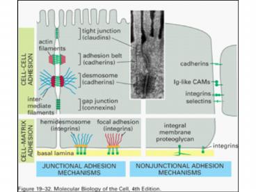
Integrins and Ig like CAMs
- After rolling on endothelial cell surface b1
integrins and b2 integrins (expressed
exclusively on leukocytes) are activated and
undergo conformational change. - At the same time vascular endothelial cells
express Ig like CAMs on their surface which act
as counter receptors for leukocyte integrins. - The leukocyte integrins bind to Ig like CAMs on
endothelial cells. - This stops the rolling and induces strong
adhesion to the endothelial cell surface.
23
.
- A mutation in the b2 integrin results in LAD
characterized by recurrent bacterial infections
due to reduction in the ability of neutrophils to
adhere to venule walls and to exit from the
blood stream. The absence of this integrin is
lethal.
24
(No Transcript)
25
Transmigration
- Locally produced cytokines released as a result
of tissue injury activate the endothelium. - Adhesion molecules such as Ig like CAMs , B1 and
B2 integrins are upregulated. - Chemoattractants such as fMLP released by
bacteria induce leukcocyte migration.
26
.
- Migration through the basal lamina requires the
activation of various digestive enzymes. - Once through the basal lamina the neutrophils
migrate up a concentration gradient of fMLP
released by bacteria. A cell can detect very
slight differences in the concentration of this
peptide in different regions of the cell surface. - These peptides are detected by receptors on the
surface of neutrophils and their occupancy by the
formyl peptides polarizes the neutrophil for
migration by reorganizing its microfilament
system.
27
(No Transcript)
28
Once neutrophils have migrated through the
endothelium they find bacteria and dead cells
through chemotaxis
29
(No Transcript)
30
(No Transcript)
31
.
- Similar mechanisms are thought to be involved in
migration of lymphocytes through the specialized
endothelium of lymph node capilaries and by
metastatic tumor cells to migrate out of venules
and capillaries .
32
Brucu DzianaWound Repair
- .
33
EpitelijaEpithelium
34
.
- Epithelial cells change in integrin expression
allowing them to change from strongly adherent
cells to less adhesive migrating cells better
able to adhere to and interact with the
extracellular components of the blood clot. - Blood clot is used as a provisional matrix for
migration of epithelial cells into the wound to
establish epithelial continuity. - Synthesis of macromolecules specific for basal
lamina and their assembly. - Proliferation of epithelial cells along the wound
edge to provide more cells.
35
SaistaudiConnective Tissue
36
.
- Fibroblasts become activated by growth factors
especially TGF beta released by cells in the
wounded region. - Fibroblasts migrate into the wound and synthesize
and secrete collagen the major component of
scars. - TGF beta also induces the fibroblasts to
differentiate into a more contractile type of
fibroblast(myofibroblast). - By pulling on the collagen fibers the
myofibroblasts induce wound contraction which
brings the two sides of the wound closer
together.
37
(No Transcript)
38
(No Transcript)
39
.
- The size of the scar depends on the time it takes
to repair the wound and on the amount of
hyaluronan present in the wounded area. - In newborns because the ECM has large amounts of
hyaluronan the wounds heal very rapidly and scars
are small or absent altogether. - If tension tending to pull the sides of the wound
apart is exerted on the wound more collagen is
secreted by the fibroblasts to resist this
tension and scars formed tend to be larger. - Once the mechanical stress on the wound is
relieved by synthesis of collagen and wound
contraction, fibroblasts in the wounded area
become quiescent and stop proliferating.
40
MetastazeanasMetastasis
- .
41
- Cancer cells
- Are genetically unstable
- Disregard signals that regulate cell
proliferation. - Avoid suicide by apoptosis.
- Avoid differentiation which normally limits
proliferation. - Escape from primary tumor.(invasive)
- Can survive and proliferate in foreign
sites.(metastasize)
42
- Normally cells recognize and adhere to
neighbouring cells by cell-cell junctions and
cell- cell adhesion molecules. - This recognition- adhesion system breaks down in
cancer. - Moreover normally cells which fail to adhere are
destroyed by apoptosis.This mechanism is also
defective in tumor cells. - This allows the tumor cells to migrate and form
metastasis.This requires changes in cell-cell and
cell -substratum adhesiveness.
43
Steps in Metastasis
- Carcinoma cells loose adhesiveness to each other.
- Overcome restraints imposed by the basal lamina
- Migrate through connective tissue.
- Penetrate blood vessels.(basal lamina)
- Migrate out of blood vessels(basal lamina)
- Migrate through connective tissue and establish a
metastasis.
44
(No Transcript)
45
(No Transcript)
46
(No Transcript)
47
(No Transcript)
48
(No Transcript)
49
Cadherins
- Cadherins and associated molecules are important
in oncogenesis and metastasis. - E-Cadherin is down regulated in most carcinomas
and is therefore a tumor suppressor. - The greater the loss of E-cadherin the greater
the metastatic potential of the carcinoma. - Loss of cadherin is accompanied by a loss of
zonula adherens junctions and a dramatic
reduction in cell-cell adhesion. - Because zonula adherens is necessary for the
maintenance of tight junctions ,they are also
disrupted resulting in the loss of cell polarity.
50
Cadherins
- Experimentally increasing the levels of
E-cadherin can restore many of the normal
epithelial properties of carcinoma cells
including loss of their ability to cause tumors
when injected into animals. - In a family in New Zealand 25 members have died
from stomach cancer.All of these individuals
carried a mutation in the gene for E-cadherin.
51
Cadherins
- Down regulation of cadherins also occurs normally
when cells separate and migrate away from the
ectoderm. - Fibroblasts induced to express E-cadherin
resemble epihelial cells.
52
(No Transcript)
53
Beta-Catenin
- When dephosphorylated on its tyrosine beta
catenin is located in zonula adherens junction
and participates in cell-cell adhesion. - Upon phosphorylation it can leave this site and
move into the cytosol or into the nucleus. - In the nucleus it can act as a transcription
factor for genes that stimulate cell replication.
54
Beta-Catenin
- In the cytosol beta-catenin is bound by APC
(Adematous polyposis coli) a protein coded by a
tumor supressor gene. - Normally the Beta catenin APC complex is then
ubiquinated and degraded. - Defects in gene coding for APC greatly increase
the risk of colon cancer. - This is likely because cytosolic beta-catenin is
not removed and is free to stimulate cell
proliferation after moving to the nucleus. - A high incidence of carcinomas is also found in
families with mutations in the beta -catenin.
55
Adenomatous Polyposis Coli
56
(No Transcript)
57
(No Transcript)
58
(No Transcript)
59
.
- To break through the basal lamina cancer cells
first upregulate integrins allowing them to
adhere strongly to laminin in the basal lamina. - They then secrete enzymes or activate enzymes (
e.g.metalloproteases ) in the ECM that digest the
basal lamina allowing the cancer cells to pass
through. Similar mechanism may allow them to pass
through the basal laminas of blood vessels. - Enzyme inhibitors could interfere with these
stages.
60
Degradation/Turnover of ECM components
Transfection of DNA coding for a defective MPP
might inhibit tumor cell metastasis
Matrix Metalloproteases FunctionTurnover,
Reorganization of ECM Cell Migration Activity
of MMPs restricted to local sites by limiting the
actvating metalloproteases to the cell surface of
the cell that needs to migrate.
61
.
- In the blood vessels the metastizing carcinoma
cells must also slow down before they can
extravasate - They probably use mechanisms similar to those
used by leukocytes,an integrin present on some
carcinoma cells that can mediate this has been
identified. - Alternately the tumor cells could become stuck in
the smaller blood vessels and then migrate out.
62
(No Transcript)
63
.
- Most tumor cells then use integrins to migrate on
fibronectin after penetrating the basal lamina. - They also use proteases to help them move through
the connective tissue. - Attempts are being made to use peptides with the
RGD sequence to inhibit these stages in
formation of metasasis.
64
Fibronectin
65
(No Transcript)
66
(No Transcript)

