Powerpoint template for scientific posters Swarthmore College
1 / 1
Title:
Powerpoint template for scientific posters Swarthmore College
Description:
Diffuse optical fluorescence imaging is based on the use of ... Fr d ric Leblond, Nicolas Robitaille, Simon Fortier, Niculae Mincu, Jean Brunette, Mario Khayat ... – PowerPoint PPT presentation
Number of Views:663
Avg rating:3.0/5.0
Title: Powerpoint template for scientific posters Swarthmore College
1
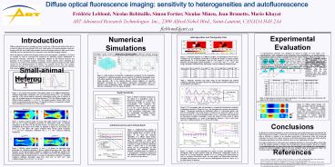
Diffuse optical fluorescence imaging sensitivity
to heterogeneities and autofluorescence Frédéric
Leblond, Nicolas Robitaille, Simon Fortier,
Niculae Mincu, Jean Brunette, Mario Khayat ART
Advanced Research Technologies Inc., 2300
Alfred-Nobel Blvd., Saint-Laurent, CANADA H4S
2A4 fleblond_at_art.ca
Experimental Evaluation A heterogeneous phantom
was designed as shown in Figure 10 (see Table 2
for specifications). Four cylindrical Cy5.5
inclusions were embedded in an autofluorescing
polyurethane matrix in order to simulate
fluorescent regions inside a diffusive medium. CW
illumination of the sample was performed at 660
nm with 1 mm resolution along both x and y axes
with a detection configuration as shown in Figure
1a). The maximum laser power available during the
scans was 38 mW. Two data sets were acquired
using different filter configurations (1)
fluorescence data (collection at a wavelength
close to the peak of the emission spectrum of the
fluorophore), (2) excitation data (collection
close to the peak of the excitation spectrum).
The resulting images are shown in Figure 11.
Numerical Simulations Simulation results consist
of optical data generated with the NIRFAST
software package 1,2. This software uses a
finite-elements method (FEM) to find numerical
solutions to the diffusion equation as well as
for more complicated cases involving fluorescent
sources. Although limited to the diffusive light
transport regime, NIRFAST is general in that it
allows the generation of solutions associated
with heterogeneous media having optical contrast
in absorption and reduced scattering, as well as
in fluorophore lifetime and concentration.
Introduction Diffuse optical fluorescence imaging
is based on the use of fluorescent probes that
can be excited by light in the near-infrared
(NIR) and visible parts of the electromagnetic
spectrum. Imaging with NIR and visible light
presents several advantages related to their
non-invasive character and low operational cost
compared to more standard imaging methods.
When designing a small-animal imaging system
having tomographic capabilities, it is critical
to evaluate as well as maximize the sensitivity
for detecting specific distributions of
fluorescent molecules. Sensitivity is
significantly affected by the internal structure
and geometry of the biological sample of
interest. Another intrinsic factor impacting the
sensitivity of a system is the ubiquitous
degradation of the signal-to-background ratio
(SBR) caused by the autofluorescence of tissues.
Our analysis shows how the complex anatomical
structure of a small animal and the
autofluorescence phenomenon influence data
acquired with diffuse optical fluorescence
systems.
Heterogeneities and Tomography Data
?a
?s
Detection Geometries
Figure 7 Schematic depiction of a diffusive
medium (corresponding to level 4 below)
considered for 2D simulations performed to
evaluate the impact of optical property
heterogeneities on a full tomography data set.
The sample is a slab with 20 mm thickness and
background optical properties ?a 0.02 mm-1 and
?s 1 mm-1 (left image absorption coefficient
map, right image reduced scattering coefficient
map).
Figure 10 Schematic showing the structure of the
heterogeneous phantom using three orthogonal
projections. The red cylinders (Fluo) represent
Cy5.5 fluorescent inclusions while the blue
cylinders (Diff) are non-fluorescent but have
optical properties different from that of the
main matrix.
Small-animal Heterogeneities
Figure 4 Light emission and detection
configurations considered for the simulations.
a) Example of a detection geometry used to
perform 3D simulated tomography scans with signal
acquisition in transmission mode. The detectors
are arranged in a cross-shape geometry with 4 mm
spacing between closest neighbors, b) Detection
geometries considered for 2D simulations
transmission configuration (left) and reflection
mode configuration (right). Tomography data is
generated by scanning a detection configuration
over the region-of-interest.
Laser
Table 1 Minimum, maximum and range values for
the absorption and reduced scattering
coefficients associated with the four
heterogeneous media considered in the 2D
simulations (see Figure 7).
Source
disease
disease
Detector
a)
b)
Figure 1 We consider data acquired with systems
based on two different illumination-collection
technologies continuous wave (CW) and
time-resolved (TR). (a) Schematic depiction of
the sourcedetector geometry (transmission mode)
used to perform a scan, (b) simulated temporal
point-spread functions (TPSF) for diffusive
samples with different optical properties (?s
1 mm-1). The impulse response function (IRF) of
the system is represented by the red curve. CW
signals (not shown) correspond to straight lines
extending over the full 12.5 ns time-window.
Normalization
Depth Sensitivity
Figure 5 Relative sensitivity (measured as CW
signal on a logarithmic scale) to fluorescent
molecules as a function of center-of-mass depth,
for various molar concentrations. The molecules
are distributed within a sphere with a radius of
1.5 mm. Curves showing a flatter response to
depth variations are related to the transmission
configuration while the exponentially decreasing
behavior characterizes the reflection mode
collection geometry.
Table 2 Heterogeneous phantom technical
specifications. The spatial locations are
referenced with respect to the origin (0,0,0)
shown in Figure 10.
Figure 11 Diffuse optical images (CW signal)
associated with the heterogeneous phantom a)
fluorescent image, b) excitation image, c)
Born-normalized image. The Cy5.5 inclusions can
only be resolved on the normalized images where
the impact of optical properties heterogeneities
is seen to be partly de-convolved.
Figure 8 CW tomography data set generated by
scanning the transmission detection configuration
depicted in Figure 4b) over the heterogeneous
samples represented in Figure 7. Fluorescent
molecules are distributed within a 1.5 mm-radius
sphere with 100 nM concentration. The
center-of-mass is located 5 mm under the surface
a) raw fluorescence data (IE), b) Born-normalized
data (IN) 3. IX represents data acquired at the
fluorophore excitation wavelength. Curves are
shown for three levels of optical property
heterogeneities, i.e., levels 0 (homogeneous
medium), 2 and 4. Each peak represents a
different detector (d1 to d5 from left to right).
Mean time (ns)
Intensity (a.u.)
Figure 2 Images produced using TR mouse data
(light emission and collection at 690 nm) for
co-axial acquisition performed in transmission
mode a) intensity (integration of the TPSF over
the full time-window), b) mean time of photon
arrival (normalized first moment of the TPSF). As
depicted in Figure 1a), the animal was placed in
a tank filled with optical matching liquid having
optical properties ?a 0.03 mm-1 (absorption
coefficient) and ?s 1 mm-1 (reduced scattering
coefficient).
- Conclusions
- Based on numerical simulations, we have shown
how non-specific fluorescence can degrade the SBR
in fluorescence images. In particular, we found
that the sensitivity of imaging devices acquiring
data in reflection is superior to an acquisition
performed in transmission when the fluorescent
molecules are close to the surface. However, we
find that transmission systems are better suited
for tomography applications since their
sensitivity does not vary significantly with
depth. - An approximation that is often made when
designing algorithms for processing diffuse
optical data is that the biological samples of
interest have homogeneous optical properties.
Based on synthetic and experimental data, we have
shown how this assumption can degrade the
correspondence between an actual tomography data
set and one computed using a homogeneous model.
We provided evidence that data normalization can
significantly improve on this situation.
Autofluorescence and Contrast-detail
Figure 6 Autofluorescence consists of photons
emanating from unlabeled tissues and/or ingested
food. As shown on the figure, it can limit the
detection capabilities of a diffuse optical
imager. Oils, pigments and proteins endogenous to
mice all contribute to whole body
autofluorescence. The figure shows SBR as a
function of depth (molecules distributed within a
sphere with a 1.5 mm-radius having 100 nM
concentration) for both transmission and
reflection detection channels. Autofluorescence
is modeled as a homogeneous fluorescent
background with different concentration levels
(curves for 1 nM and 5 nM are shown).
AAV ()
?s(mm-1)
?a (mm-1)
Figure 3 Effective optical properties (?a and
?s) of tissues are important input parameters
for algorithms designed to process optical
fluorescence data. Given light transport can be
approximately modeled by solutions to the
diffusion equation, the optical properties can be
determined by curve fitting of the TPSFs. The
images show the effective optical contrast
present in a mouse a) absorption coefficient, b)
reduced scattering coefficient. Absorption varies
from 0.015 mm-1 to 0.097 mm-1 while scattering
ranges from 0.81 mm-1 to 1.88 mm-1.
References 1 S. C. Davis, H. Dehghani, J.
Wang, S. Jiang, B. W. Pogue, K. D. Paulsen,
Image-guided diffuse optical fluorescence
tomography implemented with Laplacian-type
regularization, Opt. Express 15, 4066-4083
(2007). 2 S. C. Davis, B. W. Pogue, H.
Dehghani, K. D. Paulsen, Contrast-detail
analysis characterizing diffuse optical
fluorescence tomography image reconstruction, J.
Biomed. Opt. 10, 050501 1-3 (2005). 3 V.
Ntziachristos and R. Weissleder, Experimental
three-dimensional fluorescence reconstruction of
diffuse media by use of a normalized Born
approximation, Opt. Lett. 12, 893-895 (2001).
b)
a)
Figure 9 Figures of merit illustrating the
impact of Born normalization 3 on tomography
data for light collection using the transmission
configuration shown on Figure 4b) a) average
amplitude variation (in ), b) average peak
displacement (in mm) as a function of inclusion
depth. On each graph, raw fluorescence and
Born-normalized fluorescence curves are shown for
heterogeneous levels of 2 and 4. Nd is the total
number of detectors.


