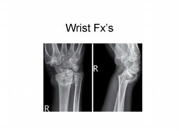
Wrist Fxs - PowerPoint PPT Presentation
1 / 32
Title:
Wrist Fxs
Description:
... and pronated wrist increasing carpal compression force on the ... Carpal displacement distinguishes this fracture from a Smith's or a Colles' fracture ... – PowerPoint PPT presentation
Number of Views:252
Avg rating:3.0/5.0
Title: Wrist Fxs
1
Wrist Fxs
2
Forearm
3
Carpals
Proximal AScaphoid, BLunate, CTriquetrum
(Triangular), DPisiform Distal ETrapezium,
FTrapezoid, GCapitate, HHamate
4
Volar Ligaments
5
Dorsal Ligaments
6
X-Ray
- Know the normal Anatomy
7
The pediatric pt
8
Suspected Fxs need 2 views
Lateral
- P/A
9
Lateral View
- Discussion
- - view should demonstrate the metacarpals,
lunate and radius aligned - - hand will appear slightly palmar flexed
10
P/A view
2mm
- On a correctly positioned PA view the extensor
carpi ulnaris tendon groove (arrow) can be seen. - The extensor carpi ulnaris tendon groove should
be at the level of or radial to the base of the
ulnar styloid.
11
Describing wrist fxs
- Location?
- Extra- or intra- articular
- most important determinant of outcome
- articular step off (only with dislocation/fx)
- second most determinant of outcome
- - radial shortening
- radial tilt dorsal or volar?
- -angulation or inclination
- radialward or ulnarward
12
(No Transcript)
13
Müller AO-classification
Adapted by the Orthopaedic Trauma Association
Colles
- A extra-articular fracture
- A1 ulna, radius intact
- A2 radius, simple and impacted
- A3 radius, multifragmentary
B partial articular fracture B1 radius,
sagittal B2 radius, frontal, dorsal rim
B3 radius, frontal, volar rim
14
Müller AO-classification
- C complete articular fracture of radius
- C1 articular simple, metaphyseal simple
- C2 articular simple, metaphyseal
multifragmentary - C3 articular multifragmentary
15
Describing wrist fxs
- Location?
- Extra- or intra- articular
- most important determinant of outcome
- articular step off (only with dislocation/fx)
- second most determinant of outcome
- - radial shortening
- radial tilt dorsal or volar?
- -angulation or inclination
- radialward or ulnarward
16
What is articular step off?
The lunate bone dislocates, or steps off the
articular surface. Often seen in Barton
Fxs Cartilage damage. Doesnt heal
wellgtgtOsteophytes
17
Describing wrist fxs
- Location?
- Extra- or intra- articular
- most important determinant of outcome
- articular step off (only with dislocation/fx)
- second most determinant of outcome
- - radial shortening
- radial tilt dorsal or volar?
- -angulation or inclination
- radialward or ulnarward
18
Lateral view Radial Tilt
- Normal volar tilt average 11 (range of 2-20).
- Determining the tilt Angle between a line along
the distal radial articular surface and the line
perpendicular to the longitudinal axis of the
radius at the joint margin. - Significance If too increased cant set fx in a
cast
19
P/A view Radial Angle (inclination)
1
3
2
- Radial inclination or angle (Average 20 degrees)
- The angle between a line connecting the radial
styloid tip and the ulnar aspect of the distal
radius a 2nd line perpendicular to the
longitudinal axis of the radius (start at distal
RA joint). - Loss of radial inclination will increase the load
across the lunate. - Cheat 2mm between the radius and lunate
- More Dislocation
- Less Impaction
20
Fx Eponyms
- Fall on an outstretched hand
- Colles
- Smith
- Bartons
- Chauffers
21
Colles Fx
- Hx Fall on an outstretched hand that is in
extension.
22
Colles Fx Displacement
Radial Tilt Dorsal Radial Length
Shortened Radial Angle Ulnar ward, Radialward or
none
23
Colles Dinner Fork
The fracture almost always occurs about 1 inch
from the end of the bone.
24
Fx?
25
Smith Fx
- Hx Falling onto flexed wrists. Less common than
Colles' fractures.
26
Smith Fx Displacement
Radial Tilt displaced volarly (ventrally) Radial
Length Shortened Radial Angle Radialward,
Ulnarward or none
27
Displacement?
28
Bartons Fx
most common fx dislocation of the wrist joint
- Hx fall on an extended and pronated wrist
increasing carpal compression force on the dorsal
rim.
29
Bartons Fx
Carpal displacement distinguishes this fracture
from a Smith's or a Colles' fracture
1
2
3
- 1. Dislocation of radiocarpal joint
- 2. may extend into the wrist joint
- 3. Often occurs along with a radial styloid fx
- Either anterior (volar) or posterior (dorsal)
cortex
30
Chauffeur's Fracture Radial Styloid Fractures
- Hx most commonly occur from ligament, tension
forces sustained during ulnar deviation and
supination of the wrist
31
Chauffer Fx Displacement
- avulsed radial styloid
- - strong radiocarpal ligament
(radioscaphocapitate) - Frequently accompanied by dislocations of lunate
32
Take Home
- AO-Classification
- Descriptive, somewhat prognostic
- Aextraarticular, Bpartial, CSOL
- A2Colles Fx
- Eponyms
- Colles Extension (Dinner Fork Deformity)
- Smith Flexion
- Bartons Dislocation along with Fx (looks like
Colles and Smith) - Chauffer Avulsion Fx (due to ulnarward deviation
supination )


