GLITTER as Forensic Evidence - PowerPoint PPT Presentation
Title:
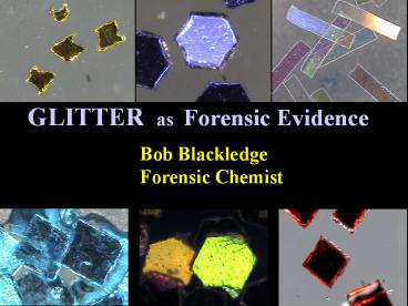
GLITTER as Forensic Evidence
Description:
GLITTER as Forensic Evidence – PowerPoint PPT presentation
Number of Views:131
Avg rating:3.0/5.0
Title: GLITTER as Forensic Evidence
1
GLITTER as Forensic Evidence
Bob Blackledge Forensic Chemist
2
Properties of the IdealContact Trace
1. Nearly invisible
2. High probability of transfer retention
3. Highly individualistic
4. Easily collected, separated, concentrated
5. Mere traces easily characterized
6. Searchable via computerized database
7. Will survive most environmental insults
3
(No Transcript)
4
Color
5
Coatings
6
Cross-section
7
(No Transcript)
8
IlluminatIR Smiths Detection, Danbury, CT
9
(No Transcript)
10
Lovedust Library Search
Lovedust
Poly(ethylene terephthalate)
11
Dispersive Raman Microspectroscopy with Confocal
Imaging
12
Raman Microspectroscopy
- The samples were thin, flat platelets and were
examined either face-on or by placing the
platelet edge-on to the Raman laser incidence for
examination of the cross-section. - A JASCO NRS-3100 Raman system fitted with 532nm
and 785nm lasers and a motorized (mapping) x-y-z
sample stage, was used. - No special sample handling was required.
- Standard microscope lenses (X5, X20 and X100)
were utilized and the X100 used for actual
measurements with a 50µm confocal aperture. - Laser powers were attenuated as appropriate to
avoid sample damage by heating, using the
build-in OD filter system of the NRS-3100.
13
Experimental conditions used
- 100mW 532nm green or 500mW 785nm deep red
solid-state lasers - 1800gr/mm (532nm) or 600gr/mm (785nm) holographic
grating - Dichroic beam splitter used with 785nm
- X100 UMPLFL objective lens
- 100nm precision automated stage
14
Amys glitter with polyester search result match
15
Crystalina 321 and 421
- The Crystalina 300 and 400 series glitters have
different polymer layer structures - To study these different layered samples we used
confocal depth mapping - The samples were probed by changing the sample
position relative to the laser spot in 1µm
increments, from the surface toward the center - The focused spot creates an effective sampling
volume of approximately 1µm diameter and 2 µm
thickness (z) - The depth resolution is therefore about 2µm
- Both Crystallina samples were confocally mapped
as 2-D (x-z) cross sections - Any metallization present was not thick enough to
affect Raman measurements - Results are shown in the following slides
16
Crystalina 421
- The 421 glitter has a clearly defined acrylic
surface layer of about 34µm on a polyester core. - There was some inevitable interference by
scattering from the adjacent layers because the
effective sampling volume z resolution is about
2µm, so for positive ID we subtracted the
adjacent layer spectrum before running the
database searches, although for the mapping, raw
peak area data were used. - The spectra, database results, and 2-D map data
follow.
17
Crystalina 421 5µm spectrum with surface spectrum
subtracted, and database search result for
polyester
18
Crystalina 421 peak 2 (PMMA) used to map layer
structure
19
Crystalina 321
- The structure of Crystalina 321 is less clear-cut
than 421 but perhaps more interesting. - The polymers appear to be mixed (copolymer or
blended). - The surface is polyester rich, with a shallow
layer slightly richer in the acrylic phase and
then becoming more polyester rich at 8-10µm
depth. - Two peak area ratios were used to follow the
layer changes in the x-z confocal map data shown
following.
20
Crystalina 321 peak area 1
21
Some case histories 1) Missouri homicide
Source
Suspects jeans
Victims bedspread
Victims jeans
22
Case 2 Illinois kidnapping sexual assault
23
Case 3 Who was driving?
24
Many thanks to
Scott Kirkowski Klaya Aardahl
Former interns/students who both now have an MFS
degree from National University, San Diego
25
Any questions?


