Case - PowerPoint PPT Presentation
1 / 61
Title:
Case
Description:
Case #1. 34 year old male with AML status-post allo-BMT complicated by severe GVHD ... Smoker with COPD, clinically like TB. Patient with known bronchiectasis ... – PowerPoint PPT presentation
Number of Views:180
Avg rating:3.0/5.0
Title: Case
1
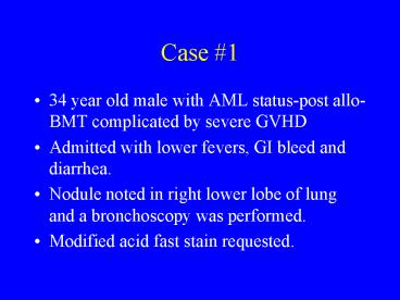
Case 1
- 34 year old male with AML status-post allo-BMT
complicated by severe GVHD - Admitted with lower fevers, GI bleed and
diarrhea. - Nodule noted in right lower lobe of lung and a
bronchoscopy was performed. - Modified acid fast stain requested.
2
BAL-Modified Acid Fast Stain
3
Case 1
4
Identification of Fungi
lt1um Branching Filaments (actinomycetes)
Filamentous fungi
Yeast
5
Actinomycetes
- Resemble fungi in that they form filaments but
their lack of mitochondria/membrane bound nucleus
and susceptibility to anti-bacterial agents
define them as bacteria. - Includes Nocardia, Nocardiopsis, Actinomadura,
Streptomyces. - Initial distinction made on basis of partial
acid-fast stain followed by chemical tests. - Nocardia are acid-fast positive.
6
Modified Acid Fast
Gram Stain
7
Nocardia
- Can cause pulmonary, systemic or cutaneous
disease. - Special request for modified acid-fast stain.
- Special request for Nocardia culture.
- Grow on Sab agar and LJ agar
- White, orange chalky colonies
- Will grow on blood, chocolate but can take over a
week and normal sputum culture plates only held
48 hours. - After suspect an actinomycetes will do further
tests to determine genus/species (e.g., lysozyme
sensitivity, casein decomposition)
8
Nocardia
9
Case 2
- 23 year old woman presents with worsening left
groin pain. - At age 15 developed cough and fever while living
in Arizona which cleared on its own. - CT scan shows retroperitoneal abscess from which
fluid was aspirated and sent for cytology and
fungal culture.
10
Case 2
11
Case 2
12
Case 2 Abscess Fluid
13
Csae 2 Abscess Fluid
14
Identification of Fungi
lt1mm Branching Filaments (actinomycetes)
Filamentous fungi
Yeast
15
Filamentous Fungi
Hyaline monomorphic
Septate hyphae
Aseptate hyphae
Saprophytes -Aspergillus -Penicillium -Fusarium -S
copulariopsis -Others
Dermatophytes -Microsporum -Trichophyton -Epidermo
phyton
Hyaline Dimorphic -Histoplasma -Coccidioides -Othe
rs
Zygomycetes -Rhizopus -Mucor -Absidia -Cunninghame
lla -Others
Dematiaceous (dark mold) -Exophiala -Clodosporiu
m -Others
16
Dimorphic Fungi
- Grow as filamentous molds in the environment or
when cultured on routine mycology agar at 25-30
degrees C - Yeast-like in vivo or when grown on enriched
media at 35-37 degrees C - Includes Histoplasma, Coccidioides,
Paracoccidioides, Blastomycoces, and Sporothrix
17
Coccidioides immitis
- Endemic in Southwestern US, Central and South
America - Typically asymptomatic or self-limiting
respiratory infection although can infect almost
any tissue. - Growth typically in 3-5 days although
characteristic arthroconidia (barrel-shaped
alternated with empty cells) may take 1-2 weeks
to grow. White cottony colony. - Appears as spherule filled with endospores in
tissue. Can produce spherules in vitro but need
special conditions and not typically done. - Infection can be confirmed by exoantigen testing
or nucleic acid probes.
18
Coccidioides-fungal form
19
Spherules and Endospores
20
Histoplasma capsulatum
Blastomycosis dermatididis
Coccidioides immitis
21
(No Transcript)
22
Case 3
- 78 yo female presented to her PCP in 1999 with
cough. Labs showed eosinophilia. CXR showed
infiltrates. AFBx3 negative. - Eosinophilia decreased but developed
bronchiectasis and pulmonary nodules. - Culture obtained from open lung biopsy.
23
MGIT System
- Middlebrook media
- Fluorescent compound on bottom that is quenched
by oxygen in the tube. - As mycobacteria consume oxygen, fluorescence can
be detected. - Most of MAC detected in nine days while MTB by 14
days. - Follow up with acid fast stain.
- Can test if fast grower by growth on MacConkey
minus crystal violet - If growth, do further chemical tests (e.g,
nitrate) - Otherwise, Gen-Probe
http//labmed.ucsf.edu/CP/SFGH/Microbiology/images
/MAI.jpeg
24
Gen-Probe
- Start with Gen-probe to MTB, MA, MI
- If negative, M. kansasii, gordonae
- If unidentified, send to State Lab
- All antibiotic testing done as send-out.
25
MAI
- Major clinical scenarios in HIV negative
patients. - Smoker with COPD, clinically like TB
- Patient with known bronchiectasis
- Elderly woman with infiltrates (Lady Windermere
Syndrome) - CT often reveals small nodules
26
Case 3 continued
- Patient presents in 2/2003 with worsened cough
and sputum production. - Sputum sent for culture.
27
Identification of Fungi
lt1mm Branching Filaments (actinomycetes)
Filamentous fungi
Yeast
28
Filamentous Fungi
Hyaline monomorphic
Septate hyphae
Aseptate hyphae
Saprophytes -Aspergillus -Penicillium -Fusarium -S
copulariopsis -Others
Dermatophytes -Microsporum -Trichophyton -Epidermo
phyton
Hyaline Dimorphic -Histoplasma -Coccidioides -Othe
rs
Zygomycetes -Rhizopus -Mucor -Absidia -Cunninghame
lla -Others
Dematiaceous (dark mold) -Exophiala -Clodosporiu
m -Others
29
Pseudallescheria boydii
30
Pseudallescheri boydiiScedosporium
apiospermumGraphium
- All the same organism
- S. apiosperum and Graphium are asexual stages.
- P. boydii is the sexual stage and you see
cleistothecia
P. boydii
Graphium
S. agiospermum
31
Pseudallescheria boydii
- Most commonly a cause of mycetoma
- Can also infect other tissues (e.g., bone, lung)
or be disseminated particularly in
immunocompromised patients. - Resistant to amphotericin B
Khurshid, A, et al., Chest (1999), 116, 572.
32
Case 4
- 55 yo male with newly diagnosed pulmonary
fibrosis. - Right middle lung biopsy showed usual
interstitial pneumonitis with active inflammatory
component c/w possible recent infections or toxic
insult. - Biopsy also sent for culture.
33
Filamentous Fungi
Hyaline monomorphic
Septate hyphae
Aseptate hyphae
Saprophytes -Aspergillus -Penicillium -Fusarium -S
copulariopsis -Others
Dermatophytes -Microsporum -Trichophyton -Epidermo
phyton
Hyaline Dimorphic -Histoplasma -Coccidioides -Othe
rs
Zygomycetes -Rhizopus -Mucor -Absidia -Cunninghame
lla -Others
Dematiaceous (dark mold) -Exophiala -Clodosporiu
m -Others
34
Aspergillus
35
Aspergillus terreus
- Cinnamon-brown colony
- Biseriate phialides, compactly columnar
- Aleuriospores found submerged in agar
36
Aspergillus niger
- Black colony
- Biseriate phialides which cover entire vesicle
and radiate
http//www.iums.org/ICPAAspnig.htm
37
Aspergillus flavus
- Light green to brown colonies. Produce
aflatoxin. - Uniseriate and biseriate phialides that cover
entire vesicle and point out in all directions. - Orange growth on Aspergillus differentiation
media.
http//www.apsnet.org/online/archive/1998/pean074.
htm
38
Aspergillus fumigatus
- Velvety, dark green colonies.
- Uniseriate phialides, usually on upper 2/3 of
vesicle. - Also can grow at 50 degrees.
39
Case 5
- Recently homeless male
- Presented to primary doctor with dystrophic
toenails and tinea pedis not responsive to OTC
anti-fungals. - Toenail sent for culture.
40
Filamentous Fungi
Hyaline monomorphic
Septate hyphae
Aseptate hyphae
Saprophytes -Aspergillus -Penicillium -Fusarium -S
copulariopsis -Others
Dermatophytes -Microsporum -Trichophyton -Epidermo
phyton
Hyaline Dimorphic -Histoplasma -Coccidioides -Othe
rs
Zygomycetes -Rhizopus -Mucor -Absidia -Cunninghame
lla -Others
Dematiaceous (dark mold) -Exophiala -Clodosporiu
m -Others
41
Scytalidium dimidiatum
- 20 year old Pakistani female rice-field worker.
- Disease began 6 years ago.
- Does not grow on mycobiotic agar (due to
cycloheximide. - 50 improvement after 6 weeks of treatment with
ketoconazole.
42
Scopulariopsis
Annellide
Conidia
- Hyaline, monomorphic, saprophyte.
- Often a contaminant but can cause nail infection.
Rarely soft tissue, bone or lungs in
immunocompromised.
43
Case 6
- 51 yo female s/p double lung transplant presents
with productive cough - CXR showed clear lungs
- Sent for flexible bronchoscopy with culture.
44
Bordetella bronchiseptica
- Gram negative pleomorphic rods.
- Unlike B. pertussis, will grow on blood and often
MacConkey. - Cause of kennel cough (dogs), snuffles (rabbits),
atrophic rhinitis in pigs. - Also know to cause pertussis like syndrome in
humans especially those exposed to animals and
immunocompromised hosts.
45
Bordetella
- Antibiotic susceptibility
- B. pertussis and parapertussis usually sensitive
to erythromycin. - B. bronchiseptica is resistant to erythromycin.
46
Case 7
- 34 yo male with AIDS presents with dizziness,
nausea and vomiting. - Develops diarrhea while in hospital.
47
Microsporidium
Trichrome stain
48
Microsporidium
Legend Pt Portion of coiled polar tubuleEx
Electron dense exosporeEn Electron lucent
endosporePv Posterior vacuole
Species by EM Treatment albendazole
http//www.zoo.utoronto.ca/dgodt/Microspridia20EM
.html
http//www.dpd.cdc.gov/dpdx/HTML/ImageLibrary/Micr
osporidiosis_il.asp?bodyM-R/Microsporidiosis/body
_Microsporidiosis_il_th.htm
49
(No Transcript)
50
E. Intestinalis vacuole
Eukaryotic cell bursting and Releasing E. hellem
spores
51
(No Transcript)
52
Case 8
- 69 yo patient found unresponsive outside after
housefire. Intubated in field, soot in airway. - Brought to BWH for burn management.
- Developed fever while in SICU. CT scan showed
free gas in peritoneum.
53
Anaerobic Isolation
Growth in anerobic blood culture bottle
Plate on CNA, KV and Brucella plates
for anaerobic growth
No growth
Gram stain. Plate on blood, chocolate and
McConkey and grow aerobically.
Gram stain, plate with kanamycin, colistin and
vancomycin discs
Gram postive, Boxcar shaped rods
Growth
Look for other features suggestive Of C.
perfringens RapidANA.
Work-up for aerobic culture.
Double zone of Beta-hemolysis
Lecthinase production
54
Clostridium perfringens
- Box-car shape
- Lecithinase positive on egg-media.
- Double zone beta-hemolysis
- Sepsis characterized by spherocytes with severe
intravascular hemolysis.
55
Case 9
- 18 yo Harvard student developed sore throat.
- Heterophile positive. Very severe sore throat
treated with steroids. - Presented one month later with SOB and pain in
right shoulder. CXR showed RLL pneumonia with
effusion (empyema). - Culture of effusion grew Streptococcus.
intermedius.
56
Case 9
- 18 yo Harvard student developed sore throat.
- Heterophile positive. Very severe sore throat
treated with steroids. - Presented one month later with SOB and pain in
right shoulder. CXR showed RLL pneumonia with
effusion (empyema). - Culture of effusion grew Streptococcus.
intermedius.
57
Strep and Staph
Streptococci in sputum sample
Staphylococci in wound sample
58
Isolation of Strep and Staph
Gram cocci
Clusters, catalase
Chains, catalase -
Staphylococcus
Streptococcus, Enterococcus
Coagulase
Coagulase -
S. aureus
R
S
Novobiocin
S. saprophyticus
S. epidermidis
59
Coagulase Test
- Initial test is Staphaurex
- Fibrinogen coated particles to detect bound
coagulase. - IgG coated particles to detect protein A
- Sensitivity 99.8
- Should do tube coagulase to detect free coagulase
if high clinical suspicion of S. aureus - Specificity 99.5
- False positives with S. saprophyticus
- Tube coagulase test (some strains only produce
free coagulase) - In plasma
- Look for clumping after 4 hours.
- To detect weak coagulase producing strains, read
again at 18 hours. Strong producers can look
negative at this point.
60
Isolation of Strep and Staph
Streptococcus, Enterococcus
Hemolysis on sheep blood agar
Alpha
Gamma
Beta
Optochin, Bile solubility
Strepstrip
Bile esculin High salt growth PYR test
Type Also, Grp A bacitracin sensitive.
Sensitive, soluble
Viridans Strep
Enterococcus
S. pneumoniae
61
Alpha-hemolytic Streptococci


