MSSBAUER SPECTROSCOPY OF CLAY MINERALS AT VARIABLE TEMPERATURES - PowerPoint PPT Presentation
1 / 1
Title:
MSSBAUER SPECTROSCOPY OF CLAY MINERALS AT VARIABLE TEMPERATURES
Description:
Geology and Geophysics, Louisiana State University, Baton Rouge, LA 70803. ... results were corrected for Compton absorption and calibrated against a-Fe foil. ... – PowerPoint PPT presentation
Number of Views:121
Avg rating:3.0/5.0
Title: MSSBAUER SPECTROSCOPY OF CLAY MINERALS AT VARIABLE TEMPERATURES
1
MÖSSBAUER SPECTROSCOPY OF CLAY MINERALS AT
VARIABLE TEMPERATURES M.D. Dyar1, E.C.Sklute1,
M.W. Schaefer2, and J.L. Bishop3. 1Dept. of
Astronomy, Mount Holyoke College, South Hadley,
MA 01075, mdyar_at_mtholyoke.edu. 2Dept. of Geology
and Geophysics, Louisiana State University, Baton
Rouge, LA 70803. 3SETI Institute/NASA-Ames
Research Ctr, Mountain View, CA 94043
Introduction Clay minerals are ubiquitous
constituents in soils on Earth, are infrequently
found in meteorites, and may also occur on
planetary surfaces in the presence of water.
However, little is known about the fundamental
Mössbauer parameters (including the recoil-free
fraction, f) that are characteristic of clay
minerals, and are critical to correctly
interpreting Fe3/SFe ratios as well as mineral
modes. Multi-temperature spectra of
well-characterized single mineral samples at
multiple temperatures are required for the
determinations of f. Thus, we present here six
layer silicates with a range of layer types.
This work is part of a larger study of these
minerals using emittance, reflectance, and
Mössbauer spectroscopies (see 1). We are also
using this study to examine the variations in
fitting results that are obtained with various
different software packages available for
processing Mössbauer data. Background This
group of minerals was chosen for our detailed
studies because they contain a range of Fe3 and
Fe2 contents and have already been
well-characterized by previous workers 2-7
(Table 1).
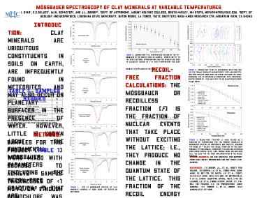
Figure 2. Isomer shift vs. temperature for one
of the Fe2 doublets in the biotite mica-Fe
sample. Points are fit to the Debye integral
approximation, and the results are used to
calculate values of f at each temperature for
each doublet as explained in the text.
Recoil-free fraction calculations The
Mössbauer or recoilless fraction (f) is the
fraction of nuclear events that take place
without exciting the lattice i.e., they produce
no change in the quantum state of the lattice.
This fraction of the recoil energy that cannot be
transferred to exciting a lattice vibration can
be quantified as f exp (-4p2ltX2gt)/l2,
where ltX2gt is the mean square vibrational
amplitude of the absorbing/transmitting nucleus
in the solid, and l is the wavelength of the g
photon. The value of f varies for different
valence states of iron in different types of
sites. The area of a Mössbauer doublet (pair
of peaks) is actually a function of peak width G,
sample saturation G(x), and the Mössbauer
recoil-free fraction f, so a correction factor
must be calculated for each doublet, as
follows where The value of f is
here determined by using the temperature
dependence (Figure 2) of the center shift (d),
which can be written as d(T) dI d SOD(T)
using the Debye model f exp
-6ER/kqD¼T/qD)2?(xdx)/(ex-1), where ER is
the recoil energy, related to the transition
energy, Eg by ER Eg2/2Mc2. Variations in
isomer shift and quadrupole splitting resulting
from use of different models Figure 3 shows a
comparison of isomer shift and quadrupole
splitting values obtained for the primary
doublets in nontronite and smectite spectra.
These results help constrain the errors in these
parameters that arise from use of varying
lineshapes. Variations in f resulting from use
of different models Values for f were
calculated at each temperature and for each
different lineshape available to us through the
three different software packages. Some of these
results are given in Figure 4. For these
samples, nearly all the fitting models give
results that yield consistent values for f. We
plan to compare these results with independent
constraints on site occupancies of Fe in order to
assess the efficacy of the Debye model.
Figure 3. Isomer shift (IS) vs. quadrupole
splitting (QS) for the primary doublets in
nontronite (top) and smectite (bottom) spectra
using eight different methods for three programs.
The IS variation is consistent with previously
quoted errors of 0.02 mm/s but the QS variation
is closer to 0.1 mm/s.
Table 1. Samples Studied
1 already determined the f values for this
sample, but we are including it in this study for
purposes of cross-comparison. Methods Samples
for this project (Table 1) were mixed with
sucrose to achieve sample thicknesses of lt1 mg
Fe/cm2 (though clinochlore was run at 5 mg
Fe/cm2). Variable temperature Mössbauer spectra
were acquired at 17 temperatures ranging from
4-295K (Figure 1). A source of 100-70 mCi 57Co
in Rh was used on a WEB Research Co. model W302
spectrometer equipped with a Janus closed-cycle
4K He refrigerator. Run times ranged from 6-24
hours, and results were corrected for Compton
absorption and calibrated against a-Fe
foil. Spectra were fit using 1) WMOSS from
WEB Research Co. in Minnesota (Lorentzian, Voigt,
and quadrupole splitting distributions, or QSD
lineshapes) 2) RECOIL from the University of
Ottawa in Canada (Lorentzian, Voigt, and QSD)
and 3) an implementation of the Wivel-Mørup
program as used at the University of Ghent in
Belgium ((Lorentzian and QSD). This resulted
in eight possible lineshape/software combinations
to use for each spectrum.
Figure 4. Recoil-free fraction (y axis) values as
a function of temperature for the primary
doublets in Mössbauer spectra of nontronite and
smectite, showing the range of f values that
result from use of the eight possible fitting
models. Errors on f values are estimated from
these curves to be 0.02, though there are
outliers (e.g., the data for the Voigt
implementation in the Recoil program.
Acknowledgments We are grateful for support from
NASA grant NNG06G130G and NSF grant
EAR-0439161. References 1 Bishop, J.L., et
al. (2007) this volume. 2 Bowen, L.H. et al.
(1989) Phys. Chem. Mins., 16, 697-703. 3 Smyth,
J.R. et al. (1997) Clays Clay Mins., 45(4)
544-550. 4 Govindaraju, K. et al. (1994)
Geostand. Newsl., 18(1), 1-42. 5 LaLonde, A.E.
et al. (1998) Hyperf. Inter., 117, 175-204. 6
Francis, C.A. (in preparation) Amer. Mineral.
7 Bishop, J., et al. (2002) Clay Minerals,
37, 617-628.
Figure 1. Fits to Mössbauer spectra of clay
minerals acquired at 295K using the DIST3E_DD
software.


