Identification of a putative transcriptional regulator - PowerPoint PPT Presentation
1 / 1
Title:
Identification of a putative transcriptional regulator
Description:
CRP TGTGATCTAGATCACA. CooA TGTCATCTGGCCGACA ... Figure 6. The alignment of recognition motifs of Crp, CooA, and Rv3676-homolog protein. ... – PowerPoint PPT presentation
Number of Views:102
Avg rating:3.0/5.0
Title: Identification of a putative transcriptional regulator
1
Identification of a putative transcriptional
regulator of CO-DH gene in Mycobacterium sp.
strain JC1 DSM 3803
JI HYANG KIM, SAE WOONG PARK, AND YOUNG M. KIM.
Department of Biology, Yonsei University, Seoul
120-749, Korea
ABSTRACT
RESULTS
Mycobacterium sp. strain JC1 is a bacterium
growing aerobically on carbon monoxide (CO) as a
sole source of carbon and energy using CO
dehydrogenase as a key enzyme. It has been
suggested that CutR may be a positive regulator
of CO-DH in this bacteria. To find other
transcription factors, proteins bound to the
putative promoter region of CO-DH structural
genes were obtained using streptavidin-agarose
column. One of these proteins was a homolog
protein of a putative transcription regulator,
Rv3676 in Mycobacterium tuberculosis H37Rv, which
belongs to the superfamily of Crp-Fnr
transcription regulators. The protein was most
closely related with CooA, which is the
transcription regulator of genes required for
CO-oxidative growth of Rhodospirillum rubrum. To
confirm the binding of Rv3676-homolog protein to
the putative CO-DH promoter region, gel mobility
shift assay was performed. Two binding complex
was formed when the purified Rv3676-homolog
protein was incubated with DNA fragments covering
the putative CO-DH promoter region. This result
suggests that the Rv3676-homolog may be another
transcription regulator involved in the
expression of CO-DH genes in Mycobacterium sp.
strain JC1.
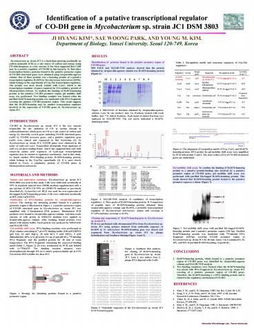
Identification of proteins bound to the
putative promoter region of CO-DH genes. SDS PAGE
and MALDI-TOF analyses showed that the protein
obtained by streptavidin-agarose column was
Rv3676-homolog protein (Figure 2).
Table 1. Recognition motifs and consensus
sequences of Crp-Fnr regulators.
Figure 2. SDS-PAGE of fractions obtained by
streptavidin-agarose column. Lane M, size marker
lane 1-6, fractions treated with wash buffer
lane 7-9, eluted fractions. Each band of eluted
fractions was analyzed by MALDI-TOF. The red
arrow indicated a Rv3676-homolog protein.
INTRODUCTION
CO-DH in Mycobacterium sp. strain JC1 is the key
enzyme responsible for the oxidation of CO to
carbon dioxide in carboxydobacteria, which grow
on CO as a sole source of carbon and energy (1).
Recently, several genes including CO-DH
structural genes (cutBCA), CO-DH accessory genes,
and a putative regulatory gene (cutR) were cloned
and sequenced in this bacterium (2). In
Mycobacterium sp. strain JC1, CO-DH genes were
clustered in the order of cutB-cutC-cutA.
Transcribed divergently from upstream of cutB was
an open reading frame. And It has suggested that
cutR with conserved amino acid sequences of
LysR-type transcriptional regulator may be
related to regulatory mechanism. And in this
study, we found another DNA-binding protein,
Rv3676-homolog protein, which belongs to the
Crp-Fnr superfamily (3). It is most closely
related to CooA, a regulatory protein of CO-DH
genes in Rhodospirillum rubrum (3).
Figure 6. The alignment of recognition motifs of
Crp, CooA, and Rv3676-homolog protein. DNA probes
for gel mobility shift assay were amplified by
PCR with primer 1 and 2. The start codon (ATG) of
CO-DH structural genes are underlined.
Gel mobility shift assay. To confirm the binding
of Rv3676-homolog protein to a putative
protein-binding sites involved by a putative
promoter region of CO-DH genes, gel mobility
shift assay was performed with purified
His-tagged Rv3676-homolog protein. This result
showed that Rv3676-homolog protein bound to the
putative promoter region as a dimer (Figure 7).
MATERIALS AND METHODS
Strains and cultivation conditions.
Mycobacterium sp. strain JC1 DSM 3803 was used in
this study. Cells were cultivated aerobically at
37? in standard mineral base (SMB) medium
supplemented with a gas mixture of 30 CO-70 air
(SMB-CO medium) as previously described (4).
Escherichia coli BL21 was used for
over-expression of his-tagged Rv3676-homolog
protein. Cells were cultivated aerobically at 30?
with 1 mM IPTG. Purification of DNA-binding
proteins by streptavidin-agarose column. The
strategy for obtaining proteins bound to a
putative promoter region is illustrated in Figure
1. A putative promoter region of CO-DH structural
gene in Mycobacterium sp. strain JC1 was
amplified with 5-biotinylated PCR primers.
Biotinylated PCR products were bound to
streptavidin-agarose column. And then crude
extracts of cells grown in SMB-CO medium were
applied to streptavidin-agarose column. After
washing, bound proteins were eluted with elution
buffer. Each protein was analyzed by SDS-PAGE and
MALDI-TOF. Gel mobility shift assay. DNA-binding
reactions were performed in 35 µl volumes
consisting of 7 µl of 5X binding buffer (100 mM
HEPES pH 8.0, 10 mM MgCl2, 30 mM KCl, 5 mM
EDTA, 5 mM dithiothreitol, 40 v/v glycerol),
0.2 µg of poly(dI?dC), 32P-labeling DNA fragment,
and the purified protein for 40 min at room
temperature. The DNA fragments containing the
conserved binding motif (Table 1, Figure 2) (5)
were synthesized by PCR and labeled with
?-32PdATP. The binding reaction mixtures were
electrophoresed through 6 (w/v) native
polyacrylamide gel in 0.5X Tris-borate-EDTA
buffer for 4h at 90 V.
Figure 3. MALDI-TOF analysis of candidates of
transcription regulators. A. Mass peaks of
Rv3676-homolog protein. B. Comparison of peptide
mass of Rv3676-homolog protein obtained from
streptavidin-agarose column and Rv3676, a
probable transcriptional regulator of
Mycobacterium tuberculosis. Amino acid coverage
is 57.14 and mass coverage is 61.9.
Cloning and sequencing of Rv3676-homolog gene
in Mycobacterium sp. strain JC1. PCR was
performed with chromosomal DNA from Mycobacterium
sp. strain JC1 using primers deduced from
nucleotide sequence of Rv3676 in M. tuberculosis.
Rv3676-homolog gene was cloned and sequenced from
Mycobacterium sp. strain JC1 by plaque
hybridization and Southern blotting (Figure 4 and
5).
Figure 7. Gel mobility shift assay with purified
His-tagged Rv3676-homolog protein and a putative
promoter region (105 bp). Purified Rv3676-homolog
protein was incubated with 32P-labeled DNA
fragments (105-bp) covering CO-DH promoter region
of Mycobacterium sp. strain JC1 for 40 min. Lanes
1 to 4 contained 0, 2X, 10X, and 20X of purified
Rv3676-homolog, respectively.
Figure 4. Southern blot analysis for cloning of
Rv3676-homolog gene in Mycobacterium sp. strain
JC1. Lane 1, size maker lane 2, phage DNA
digested with EcoRI.
CONCLUSIONS
- Rv3676-homolog protein, which bound to a putative
promoter region of CO-DH genes was identified by
streptavidin-agarose column and MALDI-TOF
analysis. - Two binding complexes were formed when the
Rv3676-homolog was mixed with DNA fragment in
Mycobacterium sp. strain JC1 covering of a
putative promoter region of CO-DH genes.
Therefore, the Rv3676-homolog is a DNA-binding
protein and is related to the regulatory
mechanism of CO-DH.
REFERENCES
- Kim, Y. M., and G. D. Hegeman. 1983. Int. Rev.
Cytol. 811-32. - Song, T. S., S. W. Park, and Y. M. Kim. 1997.
A-28, Gordon Research Conference, Henniker, NE,
USA. - Heinz K., H. J. Sofia, and W. G. Zumft. 2003.
FEMS Microbiol. Review, 27559-592. - Kim, Y. M., and G. D. Hegeman. 1981. J.
Bacteriol. 148904-911 - Shelver, D., R. L. Kerby, Y. P. He, and G. P.
Roberts. 1995. J. Bacteriol. 1772157-2163
Figure 1. Strategy for obtaining proteins bound
to a putative promoter region.
Figure 5. Nucleotide sequences of the
Mycobacterium sp. strain JC1 Rv3676-homolog gene.
Molecular Microbiology Lab., Yonsei University


