A Functional Approach to Sports Injuries - PowerPoint PPT Presentation
1 / 43
Title:
A Functional Approach to Sports Injuries
Description:
External Rotation: arm at side, elbow flexed 90 degrees. Thumb should point down to floor ... Elastic resistance exercises rotator cuff. Internal rotation ... – PowerPoint PPT presentation
Number of Views:165
Avg rating:3.0/5.0
Title: A Functional Approach to Sports Injuries
1
A Functional Approach to Sports Injuries Dr.
George R. Biltz, March 18, 2008 Childrens
Emergency Department School of Kinesiology,
University of MN
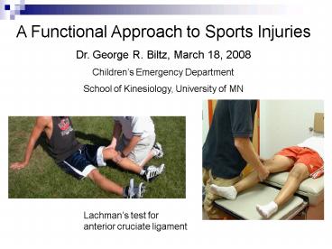
Lachmans test for anterior cruciate ligament
2
Reasons for Developing a Musculoskeletal Skill
Set
- Sports injuries accounted for 41 of all
musculoskeletal injuries treated in ED. (Ped.
Emer. Care 19(2)65-76, 2003) - Musculoskeletal problems estimated to be 10-15
of general pediatric practice. - Pediatric residency programs survey reported only
37 had hands-on teaching in Sports Medicine. (
Pediatrics 115(1) 28-33, 2005) - Chief residents reported that 29 of programs did
not include musculoskeletal examination teaching
in their curriculums. If so, it was the most
poorly taught. ( ibid )
3
A Musculoskeletal Skill Set Expands the Scope of
Your Pediatric Practice
4
Functional Orientation
- There are 3 basic questions for any sports
injury - What is the problem?
- What can be done about it?
- When can she/he return to participation?
- My goal is to help you answer those questions by
developing a functional approach to sports
injuries.
5
Whats the Problem?
- Our first goal is NOT to NAME the problem!
- Our fundamental clinical role is to manage
uncertainty - Our uncertainty about the problem
- Parent/Patient uncertainty about the problem
- Our first goal is to determine the functional
loss - thats the problem.
6
Mystery Trauma
Repetitive Microtrauma
Acute Macrotrauma
Injury Dysfunction Cycle
Adapted Manual of Sports Medicine, Eds Safran,
et al, Lippincott-Raven, 1998, p.340
Prevent or Break Cycle
Chronic Pain and Disability
7
Key Features of Functional Loss
- History of the injury and loss think
mechanically - Reported pain - location, pattern, trend
- Appearance deformity, discoloration, swelling,
inflammation - Active range of motion strength against gravity
Compare findings to the opposite side
8
Key Features of Functional Loss
- Palpable exam sites of pain, joint effusion,
swelling, heat, muscle spasm - Passive range of motion - if loss of active rom
- Weight bearing or tested strength
- Joint specific functional tests SM skill set
Compare findings to the opposite side
9
Managing the Uncertainty
- What is the best explanation for the functional
loss macro-, micro-, mystery? - Is the loss stable or progressive?
- Is the problem inside or outside of the involved
joint? ( names of potential dx?) - Will an X-ray or MRI be helpful?
- Do I need help managing the apparent uncertainty?
(mine or parents/patients)
10
Is the problem inside or outside the joint?
- Joint Effusion as a Marker for Intra-articular
Problems - loss of contour
- loss of ROM
- rapid onset suggests
- hemarthrosis
- fluid wave at knee
- patella floats in ext.
11
Is the problem inside or outside the joint?
- Upper Extremity Assessment
- Fingers and Hand flex, extend, spread fingers
make finger-thumb circle, make fist - Wrist - flex, extend, pronate supinate
forearm ( pain at either end of radius) - Elbow flex, extend (sprains are unlikely!)
- Shoulder- range of motion in 8 directions
rotator cuff - abduction, int./ext. rotation - instability - multidirectional / unidirectional
- SLAP glenoid labral tear
12
Risk Features to Recognize on Exam
Loss of Finger Tip Flexion
Rotational Deformity
13
Risk Features to Recognize on Exam
Skiers Thumb
Mallet Finger
Boutonnniere Deformity
14
Impingement Syndrome - inflammation of
supraspinatus tendon
Biceps Tendonitis
Overhead throwing motion tennis serve,
volleyball spike
15
Functional Tests at the Shoulder
Impingement Neers test (arm overhead internal
rotation) Hawkins sign (abduct, int. rotate)
Biceps Tendonitis Speeds test
16
Rotator Cuff Tendonitis abduct - supraspinatus
external rotation - infraspinatus,
teres minor internal rotation - subscapularis
17
Functional Tests at the Shoulder
Supraspinatus test
External Rotation arm at side, elbow flexed 90
degrees
Internal Rotation Lift-off test
Thumb should point down to floor
18
Functional Tests at the Shoulder
Instability Apprehension test
SLAP - superior labrum anterior posterior
OBriens test
External rotation with pressure duplicating
anterior dislocation
19
Is the problem inside or outside the joint?
- Lower Extremity Assessment
- Ankle flex, extend, ext. rotation at 90 deg.
- lateral and medial ligaments malleoli,
- Achilles tendon,
- drawer sign for stability
- Knee collateral and cruciate ligaments,
- meniscal assessment
- patella glide, tendons and attachments
- Hip flex, extend, int. and ext. rotation
20
Functional Tests at the Ankle
Syndesmosis Test
Drawer Test
21
Functional Tests at the Knee
Medial Collateral Lig.
Lateral Collateral Lig.
22
Functional Tests for Intra-articular Knee Problems
Anterior Cruciate
Meniscal compression
23
Will an X-ray be helpful?
- Assess joint effusion intra-articular injury
- articular fractures - Salter Harris II-IV, VI
- osteochondritis dessicans
- avascular necrosis Legg Perthes
- cruciate ligament avulsion
- Peri-articular pain and swelling physeal fx.
- Salter-Harris fractures I , V (compression)
- torus fx.
- tendon avulsion fx.
24
Will an X-ray be helpful?
25
Will an X-ray be helpful?
Focal pain over distal radius Salter I
Focal pain in snuff box or on volar scaphoid
tubercle scaphoid fx.
26
Elbow Epiphyseal Centers X-ray visibility order
CRITOE Capitellum, Radius, Int. epicondyle,
Trochlear, Olecranon, Ext. epicondyle
27
Risk Feature to Recognize at Elbow posterior fat
pad sign
28
Anterior Humeral Line intersects anterior 1/3 of
capitellum
29
Radiocapitellar Line radial axis aligns with
middle of capitellum
30
Helpful Shouder View - Y View for Glenohumeral
Alignment
31
Will an X-ray be helpful ?
Osteochondritis dissecans
Osgood-Schlatters
32
Will an X-ray be helpful ?
Diagnosis?
33
What can be done about it?
- Stabilization and Referral
- Staged Reassessment 4 Functional Stages
- Limit acute pain, swelling, and re-injury
- Re-establish range of motion, proprioception
- Regain strength throughout ROM
- Progress to sport specific activities
- Reassurance and Recheck if Changing
- Work-up Mystery Trauma
34
When can she/he return to participation?
- Staged Reassessment 4 Functional Stages
- Limit acute pain, swelling, and re-injury
- Re-establish range of motion, proprioception
- Regain strength throughout ROM
- Progress to sport specific activities and
demands - Recovery stages are the same but rate of
- progress varies degree of injury, individual
- Injuries do not come with expiration dates!
35
Staged Reassessment 4 Functional Stages1.
Limit pain, swelling, re-injury and dysfcn. cycle
Clinical moves 48-72 hours Ibuprofen Ice
massage Elevation Sling/splint - removeable Off
weight bearing Follow-up X-ray results
36
Staged Reassessment 4 Functional Stages2.
Re-establish range of motion and proprioception
Clinical moves 3 - 5 days Warmth (if swelling
has improved) Relax muscle tightness
(massage) Pattern and extend ROM Warmth-ROM-ice
massage repeats Partial weight bearing Isometric
muscle tension (straight leg lifts at
knee)
37
Staged Reassessment 4 Functional Stages3.
Regain strength throughout ROM
Clinical moves 5 -10 days Strength against
gravity upper extrm. Elastic resistance / small
weight Advance weight bearing lower
extrm. Non-impact activities (exercise bike) Body
weight activities of daily life Reassess
functional capacity (office)
38
3. Regain strength throughout ROM Elastic
resistance exercises rotator cuff
Internal rotation muscle?
External rotation muscles?
39
Staged Reassessment 4 Functional Stages3.
Regain strength throughout ROM
Wall Sits
Single Leg Knee Dip
40
Staged Reassessment 4 Functional Stages4.
Progress to sport specific activities
Clinical moves 10 -14 days Demonstrated
functional readiness Approve return to
conditioning Transfer follow up to team
trainer Protect joint during return Not
Improving Further evalution MRI ? Referral
Physical Therapy
41
Staged Reassessment 4 Functional Stages4.
Progress to sport specific activities
Duck Walk
Single Foot Toe Raise
42
4. Progress to Sport Specific Activities Protect
joint during return to full activity Variety of
ankle and knee braces
43
FUNCTIONALITY


