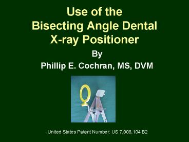
Use of the Bisecting Angle Dental Xray Positioner
1 / 61
Title: Use of the Bisecting Angle Dental Xray Positioner
1
Use of the Bisecting Angle Dental X-ray
Positioner
- By
- Phillip E. Cochran, MS, DVM
United States Patent Number US 7,008,104 B2
2
Principle of the Bisecting Angle Intraoral Dental
Radiography Imaging Technique
3
Parallel ImagingTechnique Review
The object of the parallel imaging technique is
to position the dental film as close to and as
parallel as possible to the tooth needing to be
radiographed. In the small animal mouth, this is
limited to the mandibular premolars and molars.
4
Bisecting Angle Imaging Technique
- The bisecting angle technique was developed
to achieve accurate geometric image formation in
those teeth in which a dental film cannot be
placed parallel to the tooth. These are the
canine teeth, incisors, and the maxillary
premolars and molars. The object is for the
central ray of the x-ray beam to be directed
perpendicular to an imaginary plane that bisects
the angle created between the plane of the x-ray
film and the long axis of the tooth.
5
Bisecting Angle Imaging Technique as Applied to
a Maxillary Premolar or Molar
a plane of the film b long axis of the
tooth c bisecting angle or the plane of
the bisecting angle d central ray of the
x-ray beam By directing the beam
orthagonally (perpendicular) to the bisecting
angle, the tooth will appear normal on the film,
and will measure quite close to the actual
dimensions of the tooth.
6
Bisecting Angle Imaging Technique as Applied to
an Upper Canine Tooth
a plane of the film b long axis of the
tooth c bisecting angle or the plane of
the bisecting angle d central ray of the
x-ray beam The bisecting angle imaging
technique requires the operator to direct an
invisible beam at an imaginary plane.
This requires a considerable amount of
imagination and estimation. The Bisecting Angle
Dental X-ray Positioner eliminates the guesswork
and allows for consistently reproducible,
geometrically accurate dental radiographs to be
obtained.
7
Bisecting Angle Dental Positioner as Applied to a
Maxillary Premolar or Molar
A plane of the film B long axis of the
tooth C bisecting angle or the plane of
the bisecting angle D central ray of the
x-ray beam B Metal slide that can be
moved up down, first to locate the
long axis of the tooth, and then
down and out of the way during the x-ray
exposure. Since it is parallel to
the long axis of the tooth, the
bisecting angle remains correct.
8
Bisecting Angle Dental Positioner as Applied to a
Maxillary Canine
A plane of the film B long axis of the
tooth C bisecting angle or the plane of
the bisecting angle D central ray of the
x-ray beam A Central axis of Handle
1, which is attached to the film
holder and film. The film is parallel
to the handles central axis and the
bisecting angle remains correct.
The geometry was proven in the patent
process.
The current film holder on the device is placed
such that A A overlap
9
The Bisecting Angle Dental X-ray Positioner
The Device and How To Use It
10
Bisecting Angle Positioner and its Accessory Parts
Movable metal arm for tooth axis determination
Attachment bar for film holders
Targeting ring
Film holder rods
2 Film Holder 2 Dental Film
4 Film Holder 4 Dental Film
2 CR-Computed Radiography imaging plate/film
Targeting ring holder rod
Targeting ring rod holder with rod tightening
screw
Tightening screw for movable rod holder
Handle 1
Handle 2
Bisecting angle locator
Thumb screw tightener to position angle
11
Bisecting Angle Positioner and its Accessory
Parts Side View
2 Dental film and film holder attached to device
Underside of 4 film holder with protective
rubber attached
Targeting ring and ring holder rod
4 Dental film
Targeting ring rod holder with rod tightening
screw
12
The film holder can be attached to the positioner
on its long or short axis. Note the two slits in
the film holder for the attachment.Shown here is
the film holder attached on its short axis.
Metal plate where film attaches
Two slits for attachment
4 size film holder
2 size film holder
Rubber backing
13
Film holder attached to the positioner on its
long axis using the 4 size film holder.
Two slits for attachment to film holder
14
Film sizes used in intraoral dental radiography
Size 4
Size 2
Size 0
Sizes 2 4 are the most commonly used. Dental
radiography is a non-screen technique used to
maximize detail. It is made possible by
minimizing the tube-film distance thus
concentrating the beam allowing low mAs
techniques.
15
Steps for using the dental x-ray positioner
- Step 1 Choose the proper size film for the
tooth to be radiographed. This may seem obvious,
however, on some large dogs, the standard size 2
film may not be sufficiently large for either the
upper carnassial teeth or the upper canine teeth
or you may want to image more than one tooth.
16
Step 2 Applying double backed tape to the film
holder.
There are a number of different brands and types
of double-sided tape. Shown her is the
heavy- duty 3M brand tape. I also use the
Scotch brand Removable Double Coated Tape. They
all work fine. The stickier the tape is the
longer it lasts but is harder to remove from the
holder.
I designed a number of film holders which
utilized a mechanical method to hold the film,
such as an attachment by means of raised grooved
edge, but the film would slip out and the raised
edge interfered with positioning. The
double-sided tape worked best and the film was
most secure.
17
This slide shows the tape in place with a number
2 sized dental film to be attached.
18
Step 3 Aligning the targeting ring with the
film holder.
The rod holder can be moved up and down so the
ring is aligned with the film holder. The rod
tightening screw enables the rod to rotate side
to side to also enable align- ment. The rod
should be positioned so the targeting ring will
be very close to the animals face.
Rod tightening screw
Rod holder tightening screw
Targeting ring rod holder
19
Dental Positioning Device ready for placement in
mouth
20
Using the Bisecting Angle Dental Positioner to
Radiograph the Upper Carnassial ToothStep 4
Align the movable metal arm with the tooths
longitudinal axis.
To illustrate proper alignment I will use a dog
skeleton. Place the device in the mouth behind
the carnassial tooth, and extend the movable
arm so it is aligned with the long axis of the
tooths root.
21
Make sure you are aimed at the tooth
22
Step 5 Tighten the thumb screw to lock the
angle into position
Once you have found the position of the film in
the mouth and the long axis of the tooth,
tighten the thumb screw to set the angle of the
positioner. The targeting ring is now directed
at the bisecting angle. The targeting ring is
parallel to the bisecting angle and beam is
perpendicular to it.
In a real situation, the targeting ring
demonstrated here would be too far away from the
dogs face.
23
Step 6 Retract the movable long axis
determination arm
The long axis determination arm is retracted back
to remove it from the path of the x-ray beam.
24
Step 7 Affix a tie or strap to the muzzle to
hold the positioner in place.
At this time I attach a Velcro strap or gauze tie
to hold the mouth closed to simulate a bite. The
lower teeth are protected by the rubber backing
on the film holder. In humans we are told to
bite down and hold a positioner in place.
In cats and small dogs the 2 film holder seems
to wedge itself in the mouth and will stay in
place by itself. On some short-nosed dogs and
cats I have used sandbags to hold the device
upright and in place during the exposure.
25
Step 8 Direct the tube head cone (beam
limiting device) toward the targeting ring.
Note I have use a gauze tie in this situation
to hold the mouth closed and support the
film holder to insure it stays exactly where I
want it to be. The targeting ring here is the
correct distance from the dogs face.
26
Step 4 Demonstration of the steps on a live
animal Align the movable metal arm with the
tooths longitudinal axis.
27
Step 5 Tighten the thumb screw to lock the angle
into position
Note normally I would not have the velcro tie
around the nose at this point.
28
Step 6 Retract the movable long axis
determination arm
29
Step 7 Affix a tie or strap to the muzzle to
hold the positioner in place.
30
Step 8 Direct the tube head cone (beam
limiting device) toward the targeting ring.
31
Appearance of a 90o lateral radiograph of the
upper carnassial tooth using the bisecting angle
positioning device
32
Proper tube head positioning to image the palatal
root of the upper canassial tooth
The film and holder are positioned slightly
rostral to the carnassial tooth so the image is
centered in the middle of the film.
33
Proper tube head positioning to image the palatal
root of the upper canassial tooth
34
Image obtained by prior tube head positioning
Note both rostral roots are visible. The tube
head is angled caudorostral at a 25-30o angle.
If performed rostro- caudal the roots of the
3rd premolar would overlap the 4th premolars
palatal root.
35
Increased caudorostral angulation separates the
roots better but the tooth distorts somewhat
(this is not considered unfavorable though)
36
Dental Radiography of theUpper Canine Teeth
Using the Bisecting Angle Positioner
The center opening of the long axis alignment
bar is used to approximate the position of the
canine tooths root (as it is the part of
primary concern). On the canine teeth (upper
lower) I usually have to figure out the angle
of alignment with the device off to one side,
then position it within the mouth as I need it.
The mouth gag shown here is no longer needed or
used. The bite holds the device in place.
37
Using the extended long axis alignment arm for
proper positioning on a transparent dog skull
model
38
Same process on a dog
39
The weight of the dogs head will generally keep
the positioner in place without a tie around the
muzzle. The radiograph is taken from slightly
lateral to medial (buccal to palatal) so the tip
of the canine root does not overlap and appears
medial to the first premolar tooths image.
40
Dental Radiography of theLower Canine Teeth
Using the Bisecting Angle Positioner
This techniques is almost identical to the upper
canine teeth. The animal is placed in dorsal
recumbency. The size and placement of the film
is dictated by the size of the animals mouth.
41
Using the axis determination arm to position the
device in alignment with the lower canine tooths
long axis.
42
Using the transparent skull model to demonstrate
the proper alignment
43
Similar to the upper canine teeth, the weight of
the dogs head is generally sufficient to keep
the positioner in place during the exposure.
44
Use of the Bisecting Angle Dental Positioner on a
Cat
Because of the size of the cats mouth, it is
more difficult to image but can be done. Another
problem in cats is interference with
the prominent lower rim of the orbit and
zygomatic arch. You cannot get directly next to
the tooth and must approximate the long axis of
the teeth.
45
To avoid showing the lower rim of the orbit, try
to have the film as angled as much as possible
up against the hard palate, this will make the
position of the targeting ring and x-ray beam
less vertical and more horizontal to the
patient.
46
Step 8
The long axis arm has been retracted and we are
ready to move the x-ray tube head into
place. Note that the film is angled steep to
the mouth and the beam is coming more from the
lateral than dorsal.
47
Appearance of a 90o lateral radiograph of the
upper arcade of a cat using the bisecting angle
positioning device
48
Correcting Positional Problems
This drawing illustrates the image obtained
with the correct bisecting angle
being utilized. The image is virtually the same
size as the actual tooth.
49
A 90o lateral radiograph using the Bisecting
Angle Device to obtain good geometric image
formation
This was done using a 4 film holder and 4
dental film
50
X-ray beam too vertical
If the beam is directed too vertically, or
too much from the dorsal aspect of the
patients mouth, the teeth will appear shortened
in relation to the actual size of the tooth.
51
The tube head was too dorsal
52
X-ray beam too horizontal
If the beam is directed to horizontally, or too
much from the lateral side of the patients
mouth, the teeth will appear elongated in
relation to the actual size of the teeth.
53
The tube head was positioned lateral and ventral
causing elongation of the roots
54
Use of the Bisecting Angle Positioning Device
using a Standard X-ray Machine
A standard x-ray machine tube head can be
lowered and tilted. The animals head can be
fixed into position using lead gloves or
sand- bags. The neck can be flexed or an
adjustable height table can be moved adjacent
to the x-ray table to position the animals body.
The exposure settings, due to the short tube-film
distance, are low and reduces patient exposure.
The actual settings will vary with machine, but
an mAs of 5 and kVp of 66 are in the ballpark.
We use 100S mA to take advantage of the smaller
filament size to increase sharpness of the image.
55
Aligning the Angle of the Tube Head with the
Targeting Ring
The bottom surface of the collimator is
posi- tioned parallel to the targeting ring
approx. 6 inches from the tooth. If the ring
and collimator surface are parallel, the beam is
perpendicular to the targeting ring. Since
the ring and bisecting angle are parallel, the
beam thus becomes perpendicular to the bisecting
angle also.
56
Focusing the Beam
The collimator is positioned close to the yellow
targeting ring as the photo illustrates. The
beam is collimated to the size of the film and
goes through the targeting ring and is focused
on the tooth root and dental film.
57
Using the Bisecting Angle Positioner with Direct
Digital Radiography
A clear plastic sleeve is attached to the 2 film
holder in which the DR image receptor in
inserted. The receptor, prior to insertion,
usually will have a plastic sleeve placed around
it to avoid contamination.
58
Using the Bisecting Angle Positioner with
Computed Dental Radiography
CR is easier to adapt to the Bisecting Angle
Dental Positioner because it will attach to the
2 film holder. It has the disadvantage that it
has to be removed for processing whereas DR does
not. Note the protective plastic covering over
the film on the left.
59
Final Thoughts Film
As with all new tools there is a learning curve.
It takes practice to get use to using this
device. Sometimes, on some dogs the lips can
get in the way during placing the long axis
directional bar, but you can use the bar to push
them aside. The old way of holding the film
with a forcep or taped to a tongue depressor, and
making a guess as to where the bisecting angle
is located is a thing of the past. With a little
practice you will wonder how you ever managed
without the Bisecting Angle Dental
Positioner. Phillip E. Cochran, DVM (go to next
slide for film)
60
(No Transcript)
61
To Order the J-1008 Bisecting Angle Dental X-ray
Positioner, Contact. . .
- Jorgensen Laboratories, Inc.
- www.JorVet.com
- Info_at_JorVet.com
- (800) 525-5614
- Or your local distributor of JorVet products


