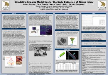
48x36 Poster Template - PowerPoint PPT Presentation
1 / 1
Title:
48x36 Poster Template
Description:
... for health care professionals in hospitals and nursing homes. ... Despite these practical attributes, there are drawbacks associated with the ultrasound. ... – PowerPoint PPT presentation
Number of Views:154
Avg rating:3.0/5.0
Title: 48x36 Poster Template
1
Simulating Imaging Modalities for the Early
Detection of Tissue Injury Kelan Hlavatya, Daria
Tanskab, Nancy Tsengb, Jia Lic, Barbara
Penprasec aCentral Michigan University, Mount
Pleasant, MI USA 48859 bUniversity of
Michigan, Ann Arbor, MI USA 48109 cOakland
University, Rochester, MI USA 48309
Imaging Technologies
Methods
Abstract
Graphs
Ultrasound is an established imaging
technique that is used in a wide variety of
medical applications. It is non-invasive and
generally accepted as a safe method. A tool that
aids in the detection of pressure ulcers using
ultrasound technology is currently being
marketed the EPISCAN I-200 produced by Longport
Inc. This device (Figure 2) is portable and can
visualize down to the bony prominence7. Despite
these practical attributes, there are drawbacks
associated with the ultrasound. The ultrasound
gel must be directly applied to the skin, which
can cause pain to the patient if tissue damage
extends to the skin surface. In addition, the
frequency range of ultrasound is limited, since
high frequencies decrease the penetration depth
and low frequencies limit the resolution of the
image13. Figure 2. The EPISCAN
I-200 shown on the left displays the images shown
on the right for a normal heel and for an
abnormal heel where blood flow is compromised.
Another imaging technique is microwave
tomography (Figure 3), which detects changes in
dielectric properties, such as relative
permittivity and conductivity9. Microwave
tomography is safe and has been used in perfusion
related problems10. However, the technology for
this technique is still premature since the
equipment is bulky and requires a complicated,
computationally expensive algorithm to decrease
the level of noise in the resulting
image10. Figure 3. The two antennas
used in microwave tomography must be carefully
placed at the sides of the affected area, as
shown here on a pigs leg. Ultra-wideband
technology is similar to microwave tomography in
that it also measures dielectric properties.
However, microwave tomography uses a narrow band
frequency while UWB operates over a wide band of
frequencies9,11. The dielectric properties
depend on the water content of the tissue, which
changes with ischemia10,11. This makes UWB an
ideal candidate for early pressure ulcer
detection. Ultra-wideband is inexpensive,
portable, and safe12. UWB transmits through
clothes and blankets, an important aspect for
patient comfort and safety12. Also featured in
UWB is an integrated transmitter and receiver12.
Thus, after considering several imaging
modalities, we decided that ultra-wideband is
most suited to test for the early detection of
pressure ulcers. To model UWB and its
sensitivity in detecting dielectric changes
associated with pressure ulcer formation, we used
two computer software programs, FEKO and XFdtd18,
to simulate the propagation of UWB waves through
a tissue model.
FEKO and XFdtd are used for electromagnetic
problems and appeared appropriate for our
purpose. FEKO uses the hybrid Method of Moments
(MoM) and Finite Element Method (FEM) technique,
which is fitting for a model with free space
between the antenna and dielectric body14. XFdtd
uses the Finite Difference Time Domain (FDTD)
method, which closely simulates the pulses of
ultra wide-band. We built a tissue model in
both programs with four layers skin,
subcutaneous fat, muscle, and bone. Each layer
was assigned the dielectric properties of
relative permittivity and conductivity these
values were found from the Italian National
Research Council15. Dielectric properties change
with frequency. In FEKO, it is possible to input
a range of frequencies, but not a range of
dielectric properties. However, XFdtd allows both
a range of frequencies and dielectric properties.
After the dielectric properties were assigned, a
plane wave was used in FEKO and a Gaussian
waveform was used in XFdtd.
Various medical imaging techniques exist to
detect the early development of tissue damage.
However, a widely commercialized device that can
be easily used and is cost-effective is still
needed. Through a literature review, we examined
ultrasound, microwave tomography, and
ultra-wideband (UWB) technology. UWB has the
capability to detect small changes in dielectric
properties, which are important since minor
alterations in perfusion and internal pressure
change dielectric properties. In addition, UWB
has the potential to become an easily accessible
technology to hospitals. Using a software called
FEKO, we attempted to simulate ultra-wideband
pulses propagating through computer-modeled
tissue layers. However, FEKO is not able to
simulate the timed pulses characteristic of UWB.
This problem is better suited for the program
XFdtd where we recreated our tissue model. Using
XFdtd, we want to show that UWB is a novel and
viable technique in detecting tissue injury.
Time Domain
Time (ps)
Graph 1. The graph on the left was generated from
FEKO and shows the near field. It is not
appropriate for analyzing changes in dielectric
properties because it encompasses only a single
frequency whereas UWB operates over multiple
frequencies. The graphs of the transmitted signal
(inset) and received signal on the right was
generated from XFdtd and is applicable for our
project because it takes into account the
frequency range of UWB.
Introduction
Horn Antenna
Tissue damage can be difficult to detect,
especially when it begins underneath the surface
of the skin. Slight changes in compartment
pressure or blood flow below the epidermis have
the potential to develop into a serious
condition, such as pressure ulcers, peripheral
vascular disease, Raynauds disease, or Buergers
disease. Advancements in imaging technology have
assisted in detecting these types of tissue
damage however, there is still a need for a more
practical device that can be easily used in a
clinical setting. We have focused on examining
pressure ulcers because of the staggering
hospital expenses associated with its formation.
Pressure ulcers are defined as a localized
area of skin and tissue injury that results from
pressure between a bony prominence and an
external surface for a prolonged period of time4.
When the bony prominence pushes on the tissue
around it, ischemia and tissue breakdown
occurs5,8. The muscle and subcutaneous fat
layers are affected first in deep tissue pressure
ulcer formation and necrosis spreads outward to
the skin5. By the time the damage is evident on
the surface of the skin, extensive injury has
already occurred to the tissue underneath5.
Advancing age, malnutrition, and diabetes are a
few risk factors for pressure ulcer formation1.
Common locations (Figure 1) are the coccyx and
heel6. Pressure ulcers are an increasing
source of concern for health care professionals
in hospitals and nursing homes. In an
eleven-year span, from 1992-2003, the number of
pressure ulcers diagnosed increased by 631.
Pressure ulcers cause pain and discomfort for the
patient and may be fatal. There were over 57,000
deaths from pressure ulcers between 2003-20052.
Additionally, substantial costs are associated
with the treatment and prevention of pressure
ulcers. In the United States, there is an
estimated 8.5 billion spent annually on pressure
ulcer treatment3. One way to diminish these costs
is through a medical imaging device focused on
early detection. Figure 1. The
diagram to the left indicates common locations
for pressure ulcers in the body. The photographs
to the right show advanced stages of a pressure
ulcer.
For the simulation of UWB propagation
through tissue layers, a modified pyramidal horn
antenna17 (Figure 4) was successfully modeled.
This horn antenna has been modified from more
expensive horn antennas since its waveguide
section has been eliminated and a curved metallic
plane has been added.
Future Work
Further research can be done with XFdtd, a
full wave electromagnetic solver based on the
Finite Difference Time Domain method, to
determine UWBs feasibility in detecting changes
in dielectric properties. Within the model, the
conductivity and permittivity values can be
adjusted to mirror a change in water (or blood)
content. The output from this modification would
show the sensitivity of UWB pulses in detecting
pressure ulcers and other types of tissue damage.
http//www.mediluxprofessional.net/pages/Medilux2
0Episcan20Brochure.pdf http//www.longportinc.com
/downloads/Acute_Care-Wound_Prevention_and_Assessm
ent.pdf
Acknowledgements
We would like to thank the SIBHI program at
Oakland University, the National Science
Foundation, and the National Institute of Health.
Li, X., et al. Numerical and Experimental
Investigation of an Ultrawideband Ridged
Pyramidal Horn Antenna with Curved Launching
Plane for Pulse Radiation." IEEE Antennas and
Wireless Propagation Letters. 2 (2003) 259-262.
Figure 4. The left shows the actual size of the
modified horn antenna, and the right shows a horn
antenna simulated in FEKO.
References
1 Elixhauser, A. and Russo, A. Statistical
Brief 3 Hospitalizations Related to Pressure
Sores, 2003. Agency for Healthcare
Research and Quality. (2006)1-7. 2 Patient
Safety in American Hospitals. Health Grades, Inc.
2004. 3 Bennett et al. The cost of pressure
ulcers in the UK. Age and Ageing. 33 (2004)
230-235. 4 NPUAP2007_PU_Def_and_Descriptions.
National Pressure Ulcer Advisory Panel. Available
at lthttp//www.npuap.org/documents/NPUAP2007
_PU_Def_and_Descriptions.pdfgt. (2007) 5 Bouten,
C. V. et al. The Etiology of Pressure Ulcers
Skin Deep or Muscle Bound? Arch Phys Med
Rehabil. 84 (2003) 616-619. 6 Cole, L. and
Nesbitt, C. A Three-Year Multiphase Pressure
Ulcer Prevalence/Incidence Study in a
Regional Referral Hospital. Ostomy/Wound
Management. 50 (2004) 32-40. 7 Acute Care
Detection, Prevention and Assessment of Pressure
Ulcers. Longport Inc. Available at
lthttp//www.longportinc.com/downloads/Acute_Care-W
ound_Prevention_and_Assessment.pdfgt. 8 Peirce,
et al. Ischemia-reperfusion injury in chronic
pressure ulcer formation a skin model in the
rat. Wound Repair and Regeneration. 8 (2000)
68-76. 9 Corfield, D. R. and Semenov, S. Y.
"Microwave Tomography for Brain Imaging
Feasibility Assessment for Stroke
Detection." International Journal of Antennas and
Propagation. (2008) 1-8. 10 Semenov, S. Y., et
al. "Microwave tomography for functional imaging
of extremity soft tissues feasibility
assessment." Phys. Med. Biol. 52 (2007)
5705-5719. 11 Lazebnik, Mariya et al. A
large-scale study of the ultrawideband microwave
dielectric properties of normal, benign
and malignant breast tissues obtained from cancer
surgeries. Physics in Medicine and
Biology. 52 (2007) 6093-6115. 12 Staderini,
Enrico M. UWB Radars in Medicine. IEEE AESS
Systems Magazine. (2002) 13-18. 13 Aspres, N,
et al. Personal Review Imaging the Skin.
Australasian Journal of Dermatology. 44
(2003) 19-27. 14 FEKO Comprehensive EM
Solutions. EM Software Systems. 16 June 2008
lthttp//www.feko.info/gt. 15 Calculation of the
Dielectric Properties of Tissue in the Frequency
Range 10 Hz - 100 GHz. Italian
National Research Council. 13 June 2008
lthttp//niremf.ifac.cnr.it/tissprop/htmlclie/htmlc
lie.htmgt. 16 Fiske, D. "New Public Safety
Applications and Broadband Internet Access Among
Uses Envisioined by FCC Authorization of
Ultra-Wideband Technology." News 14 Feb. 2002. 30
June 2008. 3 July 2008.
lthttp//http//www.fcc.gov/Bureaus/Engineering_Tec
hnology/News_Releases/2002/nret0203.pdfgt. 17
Li, X., et al. Numerical and Experimental
Investigation of an Ultrawideband Ridged
Pyramidal Horn Antenna with Curved
Launching Plane for Pulse Radiation." IEEE
Antennas and Wireless Propagation
Letters. 2 (2003) 259-262. 18 Xfdtd. Remcom. 8
July 2008 lthttp//www.remcom.com/products.htmlgt.
Tissue Model
Semenov, S. Y., et al. "Microwave tomography for
functional imaging of extremity soft tissues
feasibility assessment." Phys. Med. Biol. 52
(2007) 5705-5719.
Figure 4. The FEKO model on the left illustrates
the four tissue layers of skin, fat, muscle and
bone as well as a plane wave, near field, and
receiving antenna. The model on the right is from
XFdtd and also illustrates the four tissue layers
as well as a simulated horn antenna.
http//www.pele.com.tw/pressure_ulcers.htm http//
www.medscape.com/viewarticle/418377_7 http//www.v
isualdxhealth.com/adult/decubitusUlcer.htm
Table 1. The above chart lists the dielectric
properties of permittivity and conductivity at
their respective frequency. These properties have
been applied to the tissue models for both FEKO
and XFdtd.

