MRI parameters - PowerPoint PPT Presentation
1 / 1
Title:
MRI parameters
Description:
parallel to the cord (//) perpendicular to the cord ( ) 2. Positioning at the ... ventilated using a MRI-1 rodent ventilator (CWE, Ardmore, USA) with 1.5 ... – PowerPoint PPT presentation
Number of Views:592
Avg rating:3.0/5.0
Title: MRI parameters
1
Inserm Institut national de la santé et de la
recherche médicale
IN VIVO MONITORING OF A MOUSE SPINAL CORD INJURY
DURING THE ACUTE PHASE BY QUANTITATIVE MRI
Jean-Marie Bonny (1), Henri Haton (3),
Jean-Pierre Renou (1), Alain Privat (3), Manuel
Gaviria (2) (1) INRA STIM QuaPa, Clermont
Ferrand, France (2) Neuréva Inc., INM,
Montpellier, France (3) INSERM U583, INM,
Montpellier, France
Introduction Cascades of neurochemical processes
begin immediately after a primary lesion of the
spinal cord. Early therapeutic management is thus
crucial in terms of control of the secondary
phenomena and functional recovery. Reliable data
of the time course of these processes are
mandatory in order to optimize the molecular
targeting and the therapeutic windows of
neuroprotective pharmacological compounds. To
date, none of the latter has shown a conclusive
clinical efficacy. In the present study, high
field NMR micro-imaging (9.4 Tesla) has been used
to follow the development of a photochemical
ischemic lesion induced at thoracic level in
mice. Due to its ability to detect the early
ischemic events as well as the demyelination
phenomenon, quantitative imaging has been carried
out for measuring the apparent diffusion
coefficients (ADC) of water, parallel and
perpendicular to the spinal axis, and the spin
relaxation time T2.
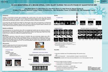
Results 4. Control mice Acquired
images 5. SCI mice
Parametric images
//
?
T2
Materials and Methods Experimental ischemic SCI
was performed using a 560 nm wavelength-light
irradiation that was carried-out in female
13-week old C57Bl/6J mice through the T8
posterior lamina. This irradiation induces the
excitation of an intraperitonealy injected
photosensitive dye, namely Rose Bengal (RB) and
provokes ischemic necrosis of the neural tissue.
After the end of the surgical procedure, animals
were conditioned for quantitative MRI
monitoring. MRI follow-up Experiments were
performed on Bruker DRX Avance 9.4T (400 MHz).
Mice were mechanically ventilated using a MRI-1
rodent ventilator (CWE, Ardmore, USA) with 1.5
isoflurane in 60 O2, 2.2-ml tidal volume, 100
breaths per min and a 11 inspiration-to-expiratio
n ratio. Fifteen minutes before intubation,
animals were atropinized with an intramuscular
25-50 µg/kg injection which was repeated every
hour. After initiation of ventilation, they were
curarized with an intraperitoneal 0.3-mg/kg bolus
of Pavulon which was repeated every 45 min. The
chronic administration of these two compounds was
done inside the magnet by means of two IM
catheters.
- Longitudinal follow-up
- important anatomical swelling of dorsal horns is
apparent on images as early as the second hour
after the lesion - other microstructural alterations are detected by
changes of quantitative MR parameters - ? decrease of ADC
Inter-animal comparison 4 hours after the injury
MRI parameters Quantitative imaging was carried
out for measuring the apparent diffusion
coefficients (ADC) of water parallel and
perpendicular to the spinal axis, and the spin
relaxation time T2 1. Acquired images -
Reference - T2 - weighted images - Diffusion -
weighted images ? parallel to the cord (//) ?
perpendicular to the cord (?) 2. Positioning
at the thoracic level T7/T8 Volume of the
voxel 0.125 x 0.125 x 1 mm3 FOV 12 x 12 mm2
Birdcage radiofrequency coil (Ø20mm), excitation
reception
- Key points
- Inter-group variability due to the
pathophysiology of the ischemic injury - but
- Reproducible approach ? robust quantitative
changes in injured territories - Results are consistent with those previously
obtained ex vivo on fixed samples in our lab
3. Minimisation of the motion artefacts
.
- Perspectives
- Procedure for analysis of groups (in progress)
- Common pattern of changes depending on the
analyzed SC region - Confirming the ex vivo characterization
- Spatial quantitative characterization ?
normative data - Screening of new potential neuroprotective
compounds - Fine tuning of pre-clinical therapeutic windows
- Clinical translation
References Bonny JM, et al. 2004 Neurobiol.Dis.
15474-482. Gaviria M, et al, 2006 Neurobiol.
Dis. 22694-701. Gaviria et al. 2002 J.
Neurotrauma, 19205-221. Song SK, et al. 2003
Neuroimage 201714-1722. Franconi F, et al.
2000 Magn.Reson.Med. 44893-898.

