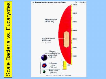
Scale Bacteria vs. Eucaryotes
1 / 50
Title: Scale Bacteria vs. Eucaryotes
1
Scale Bacteria vs. Eucaryotes
2
Bacteria vs. Eucaryotes
3
Three Cellular Domains
16S rRNA
4
Three Cellular Domains
5
Ribosome Image
6
Diversity of Bacterial Shapes
7
Coccus (pl. Cocci)
8
Diversity of Bacterial Shapes
9
Coccobacillus
10
Diversity of Bacterial Shapes
11
Vibrio
12
Diversity of Bacterial Shapes
13
Bacillus (pl. Bacilli)
14
Escherichia coli
15
Diversity of Bacterial Shapes
16
Spirillum
17
Diversity of Bacterial Shapes
18
Spirochete
19
Bacterial Cellular Arrangements
20
Filamentous Bacteria
21
Bacterial Anatomy (Overview)
22
Nucleoid
23
Endospores
24
Bacterial Anatomy (Cell Envelopes)
25
Gram Positive Cell Envelope
26
Gram Negative Cell Envelope
27
Lipopolysaccharide
28
Endosymbiosis
29
Mitochondrian
30
Chloroplast
31
Bacterial Anatomy (Plasma Membrane)
32
Plasma Membrane
33
Movement Across Membranes
water
34
Osmosis Tonicity
Cell Walls
Plasmolysis
35
Protoplast Spheroplast
A Protoplast is a Gram-Positive bacterium without
its cell wall
A Spheroplast is a Gram-Negative bacterium
lacking most of its cell wall
36
Bacterial Anatomy (Flagella)
37
Flagellum (p. Flagella) (1/2)
also atrichous
38
Flagellum (p. Flagella) (2/2)
39
Taxis
Negative Chemotaxis is away from specific
substances
Positive Chemotaxis is towards specific substances
Negative Phototaxis is away from light
Positive Phototaxis is towards light
40
Axial Filament (EndoFlagella)
41
Bacterial Anatomy (Pili)
42
Pilus (pl. pili) -- Fimbria
43
Sex Pili
44
Cell-Surface Fibrils
Electron micrograph of an ultra-thin section of a
chain of group A streptococci. The cell surface
fibrils, consisting primarily of M protein, are
clearly evident. The bacterial cell wall, to
which the fibrils are attached, is also clearly
seen as the light staining region between the
fibrils and the dark staining cell interior.
Incipient cell division is also indicated by the
nascent septum formation (seen as an indentation
of the cell wall) near the cell equator. The
streptococcal cell diameter is equal to
approximately one micron.
45
Bacterial Anatomy (Glycocalyx)
46
Glycocalyx
47
Bacterial Anatomy (Overview)
48
Link to Next Presentation
49
(No Transcript)
50
Laboratory Primer
Just reading a lab exercise is not the same as
getting ready to do a labyou also need to
outline for yourself, either mentally or on
paper, just what it is that you will be doing
I know that making such an outline with
unfamiliar material is not easythat is why you
need to look at your lab schedule, where I
attempt to guide you through what it is that you
will need to be doing
You have to try to remember that a culture that
has settled will need to be resuspendedand you
have to not just go through the motions you
actually need to resuspend it!
It may be that some of you have not had previous
training in using a microscope (though isnt it
part of bio 101?) ? after class today we will
have a microscope 101 session in B211


