Chapter 16 Lymphatic System - PowerPoint PPT Presentation
Title:
Chapter 16 Lymphatic System
Description:
Chapter 16 Lymphatic System – PowerPoint PPT presentation
Number of Views:81
Avg rating:3.0/5.0
Title: Chapter 16 Lymphatic System
1
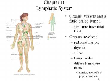
Chapter 16Lymphatic System
- Organs, vessels and a fluid called lymph
- similar to interstitial fluid
- Organs involved
- red bone marrow
- thymus
- spleen
- lymph nodes
- diffuse lymphatic tissue
- tonsils, adenoids peyers patches
2
Functions of the Lymphatic System
- Draining excess interstitial fluid plasma
proteins from tissue spaces - Transporting dietary lipids vitamins from GI
tract to the blood - Facilitating immune responses
- recognize microbes or abnormal cells responding
by killing them directly or secreting antibodies
that cause their destruction
3
Lymphatic Vessels Circulation
- Capillaries that begin asclosed-ended tubes
foundin spaces between cells - Combine to form lymphaticvessels
- resemble veins with thinwalls more valves
- Fluid flows through lymph nodes towards large
veins above the heart - lymph emptied into bloodstream
4
Lymphatic Capillaries
- Found throughout thebody except in
Avasculartissue (cartilage, epidermis cornea) - Structure is designed to lettissue fluid in but
not out - anchoring filaments keep tubefrom collapsing
under outside pressure - overlapping endothelial cells open when tissue
pressure is high (one-way valve) - In GI tract, known as lacteals -- contain chyle
5
Lymph Trunks Ducts
- Vessels unite to form trunks thoracic ducts
- Right side head, arm chest empty into right
lymphatic duct and rest of body empties into
thoracic duct - Lymph is dumped directly into left right
subclavian veins
6
Formation Flow of Lymph
- Fluid proteins escaping from vascular
capillaries is collected by lymphatic capillaries
returned to the blood - Respiratory muscular pumps promote flow of
lymphatic fluid - Lymphatic vessels empty into subclavian veins
7
Lymphatic Organs Tissues
- Widely distributed throughout the body
- Primary lymphatic organs
- provide environment for stem cells to divide
mature into B and T lymphocytes - red bone marrow gives rise to mature B cells
- thymus is site where pre-T cells from red marrow
mature - Secondary lymphatic organs tissues
- site where most immune responses occur
- lymph nodes, spleen lymphatic nodules
8
Thymus Gland
- Large organ in infants (70 g) but atrophied as
adult (3 g) - 2 lobed organ located in mediastinum
- Capsule trabeculae divideit into lobules
- Each lobule has cortex medulla
- Cortex
- tightly packed lymphocytes macrophages
- Medulla
- reticular epithelial cells produces thymic
hormones - Hassalls corpuscles
9
Lymph Nodes
- Flow is in one direction
- afferent vessels lead in
- sinuses lead to efferent vessels that exit at
hilus - Only nodes filter lymph
10
(No Transcript)
11
Lymph Nodes
- Bean-shaped organs, up to 1 inch long, located
along lymphatic vessels - scattered throughout body but concentrated near
mammary glands, axillae groin - Stroma is capsule, trabeculae reticular fibers
- Parenchyma is divided into 2 regions
- cortex
- lymphatic nodules with germinal centers
containing dendritic cells - antigen-presenting cells and macrophages
- B cells proliferate into antibody-secreting
plasma cells - medulla
- contains B cells plasma cells in medullary cords
12
Spleen
- 5 inch organ between stomach diaphragm
- Hilus contains blood lymphatic vessels
- Stroma consists of capsule, trabeculae, fibers
fibroblasts - Parenchyma consists of white pulp and red pulp
- white is lymphatic tissue (lymphocytes
macrophages) around branches of splenic artery - red pulp is venous sinuses filled with blood
splenic tissue (splenic cords)
13
Lymphatic Nodules
- Concentrations of lymphatic tissue not surrounded
by a capsule scattered throughout connective
tissue of mucous membranes - mucosa-associated lymphoid tissue (MALT)
- Peyers patches in the ileum of the small
intestine - Appendix
- Tonsils form ring at top of throat
- adenoids (pharyngeal tonsil)
- palatine tonsils (on each side wall)
- lingual tonsil in the back of the tongue
14
Principal groups of lymph nodes
15
(No Transcript)
16
(No Transcript)
17
(No Transcript)
18
(No Transcript)
19
(No Transcript)

