STRESS FRACTURES - PowerPoint PPT Presentation
1 / 45
Title:
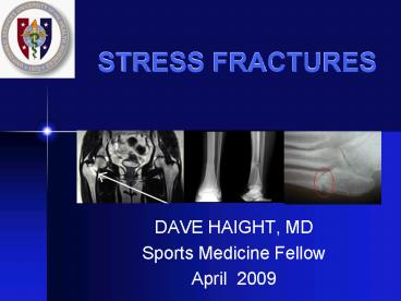
STRESS FRACTURES
Description:
Seen more commonly in jumpers and leapers. See 'dreaded black line' on x-ray. Heal very poorly ... Consider in: Sprinters, Jumpers, Hurdlers, Basketball, Football ... – PowerPoint PPT presentation
Number of Views:959
Avg rating:3.0/5.0
Title: STRESS FRACTURES
1
STRESS FRACTURES
- DAVE HAIGHT, MD
- Sports Medicine Fellow
- April 2009
2
Outline
- Pathophysiology
- Risk Factors
- Associations
- Diagnosis
- General Treatment
- Specific Cases
3
CAUSE
- Change in load
- Small number of repetitions with large load
- Large number of reps, usual load
- Intermediate combination of increased load and
repetition
4
PATHOPHYSIOLOGY
- Wolffs Law change in external stress leads to
change in shape and strength of bone - bone re-models in response to stress
- ABRUPT Increase in duration, intensity, frequency
without adequate rest (re-modeling) - Stress fracture imbalance between bone
resorption and formation - Microfracture -gt continued load -gt stress fracture
5
EPIDEMIOLOGY
- 1 of general population
- 1-8 of collegiate team sports
- Up to 31 of military recruits
- 13-52 of runners
6
RISK FACTORS
- History of prior stress fracture
- Low level of physical fitness, non-athlete
- Increasing volume and intensity
- Female Gender
- Menstrual irregularity
- Diet poor in calcium
- Poor bone health
- Poor biomechanics
7
RISK FACTORS cont
- Prior stress fracture
- 6 x risk in distance runner and military recruits
- 60 of track athletes have hx of prior stress
fracture - One year recurrence 13
- Poor Physical Fitness - muscles absorb impact
- gt1cm decrease in calf girth
- Less lean mass in LE
- Less than 7 months prior strength training
8
INTRINSIC FACTORS
- Extreme arch morphologies
- Pes cavus
- Pes planus
- Biomechanical factors
- Shorter duration of foot pronation
- Sub-talar joint control
- Tibial striking torque
- Early hindfoot eversion
9
EXTRINSIC FACTORS
- Activity type and intensity
- Footwear
- Older shoes
- Shock absorbing cushioned inserts
- Running Surface
- Treadmill
- Track
10
ASSOCIATIONS
- Ballet
- Runners
- Sprinters
- Long dist runner
- Baseball, tennis
- Gymnasts
- Rowers, golfers
- Hurdlers
- Rowers, Aerobics
- Bowling, running
- Lumbar, femur, metatarsal
- Tibia, metatarsal
- Navicular
- Femoral neck, pelvis
- Humerus
- Spine, foot, pelvis
- Ribs
- Patella
- Sacrum
- Pelvis
11
Classic Clinical History
- Change in training or equipment
- Gradual onset over 2 to 4 weeks
- Initially pain only with activity
- Progresses to pain after activity
- Eventually constant pain with ADLs
12
DIAGNOSISHistory
- Sports participation
- Significant change in training
- Hills, surface, intensity
- Dietary History adequacy, Vit D, Calcium
- Menstrual History
- General Health
- Occupation
- Past medical history
- Medications
- Family history (osteoporosis)
13
IMAGINGX-ray
- Only 30 positive on initial examination
- 10 - 20 never show up on plain films
- If a positive x-ray
- Localized periosteal reaction
- Radiolucent line
- Cancellous bone - band-like focal sclerosis
14
Early Metatarsal Stress Fracture
15
One Week Later..
16
Bone Scan
- 95 show up after 1 day
- Extremely sensitive but not as specific with up
to 24 false-positive results (stress reaction) - Differentiate between acute and old lesions
- Acute stress fracture three phase positive
- Shin splint delayed phase only
17
MRI vs. bone scan, CJSM 2002
- MRI less invasive, provided more information than
bone scan and recommended for initial diagnosis
and staging of stress injuries - Limited MRI may be cheaper than bone scan at
some institutions
18
How I Decide Between an MRI and Bone Scan
- MRI
- Usually can be done more quickly (1 vs. 4 hours)
and scheduled for a sooner date - No radiation
- Better soft tissue detail
- Bone Scan
- Covers a wider area of the body (if bilateral or
diffuse symptoms) - Sometimes easier to interpret
- Cheaper
19
RADIATION COMPARISON
- Study mSv relative radiation
- Plain film foot lt0.01 lt 1.5 days
- Plain film CXR 0.02 2.4 days
- Plain film pelvis 0.7 3.2 mo
- Tech-99 bone scan 3 (150 CXR) 1.2 yrs
- CT L-spine 6 (300 CXR) 2.3 yrs
- CT abd / pelvis 10 (500 CXR) 4.5 yrs
20
GENERAL TREATMENT
- PROTECTION
- Reduce pain
- Promote healing
- Prevent further bone damage
- ADLs are permitted
- Stretching and flexibility exercises
- Cross-training (non-weight-bearing exercise)
- Modified rest for six to eight weeks (or until
pain-free for two to three weeks)
21
ACTIVITY MODIFICATION
- Activity should be pain free
- Approximate desired activity
- Cycle
- Swim
- Walk
- Elliptical
- Deep water running
- MUST BE PAIN FREE
22
REHAB EXERCISE
- Address biomechanical issues
- Muscle inflexibility
- Limb Length Discrepancy
- Excessive pronation, pes cavus, pes planus
- Replace running shoes
- Strength training
23
Site of Stress Fractures
- Tibia - 39.5
- Metatarsals - 21.6
- Fibula - 12.2
- Navicular - 8.0
- Femur - 6.4
- Pelvis - 1.9
24
HIGH RISK
- High Risk
- Talus
- Tarsal navicular
- Proximal fifth metatarsal
- Great toe sesamoid
- Base of second metatarsal
- Medial malleolus
- High Risk
- Pars interarticularis
- Femoral head
- Femoral neck
- (tension side)
- Patella
- Anterior cortex of tibia
- (tension side)
25
High-Risk Tibial Stress Fx
- Anterior, middle-third stress fractures are very
concerning - Tension side of bone
- May present like shin splints
- Seen more commonly in jumpers and leapers
- See dreaded black line on x-ray
- Heal very poorly
26
Dreaded Black Line
27
Management of High-Risk Tibial Stress Fx
- Immobilization
- 4-6 months of rest
- Pulsed low-intensity U/S or electrical
stimulation may decrease symptoms and speed
return to activity, 30 minutes/day x 3-9 mos. - IM rod for failed conservative or patient
preference
28
5th metatarsal stress fracture
29
Types of Proximal 5th MT Fractures
30
Mgmt. of 5th Metatarsal Stress Fx
- High risk for delayed union or nonunion
- Non-weight-bearing cast for 6 weeks versus IM
screw fixation
31
IM Screw Fixation
32
Spondylolysis
- Stress fracture of the pars interarticularis
- Caused by repetitive hyper-extension
- Often develops in the teenage or pre-teen years
- May be bilateral
33
Sports Associated with Spondys
- Football (offensive lineman)
- Gymnastics
- Wrestling
- Diving
- Tennis
- Volleyball
34
Physical Exam- Spondy
- Tenderness to palpation over paraspinous muscles
- Positive one-legged hyperextension test
- Stork test
- Tight hamstrings- cause or effect?
35
Radiographs- Spondy
- ? Need to get oblique images
- Look for Scotty Dog sign
- SPECT scan
- MRI not reliable in diagnosing
36
SPECT scan showing bilateral pars defects
37
Treatment- Spondy
- Relative rest (avoid lunges, cleans, squats,
other extension maneuvers, etc.) - Williams flexion exercises
- Pelvic tilt, Single Knee to chest, Double knee to
chest, Partial sit-up, Hamstring stretch, Hip
Flexor stretch, Squat (no weight) - Anti-lordotic bracing
- Return to activity in brace when pain-free
- Brace 6 weeks - 6 months (controversial)
38
Femoral Stress Fx
- Primary presenting symptom is groin pain
possibly thigh or knee pain - Hip motion may be painful
- Hop test
- Fulcrum test for shaft fx
39
Femoral Neck Stress Fx
- Early diagnosis critical
- If x-rays negative, bone scan/MRI
- MRI diagnostic imaging of choice for femoral neck
stress fractures
40
Femoral Neck Stress Fx
- Compression side.
- Inferior part of femoral neck
- Younger patients
- Less likely to become displaced
- Complications possible
- Treatment-non-weight bearing, followed by
touch-down WB, then partial WB over a total of
8-12 weeks
41
Femoral Neck Stress Fx
- Distraction side
- Superior cortex or
- tension side of neck
- High propensity to become displaced
- Frequent complications
- Treated acutely with internal fixation
42
Tarsal Navicular Stress Fx
- Consider in Sprinters, Jumpers, Hurdlers,
Basketball, Football - Mean interval of 7 -12 months before diagnosis
- Vague mid-foot pain
- Pain on dorsum of foot
- Foot cramping
43
(No Transcript)
44
(No Transcript)
45
Tarsal Navicular Stress Fx
- X-rays usually negative
- Bone scan vs. MRI vs. thin cut CT
46
Mgmt. of Navicular Stress Fx
- Most studies suggest that allowance of
weight-bearing immediately after diagnosis
increases the non-union rate - General/simple rules
- () bone scan/MRI/CT and/or incomplete fx- NWB
cast x 6-8 weeks, and then gradual rehab - Complete fx and/or bony sclerosis- ORIF with
compression screw /- bone graft
47
Navicular Stress Fx- Return to Play
- After casting, if no tenderness at the N spot,
then can gradually return - Reassess every 1-2 weeks, gradual return at 6
weeks if no symptoms - AFTER 6 weeks of protection, 6 weeks of PT for
strength and flexibility prior to return to run! - Average return to play is 4-6 months
- Follow up radiography not helpful for return to
activity
48
Sesamoid stress Fx
- Risk Sudden start-stop sports
- Repetitive forced dorsiflexion
- Conservative tx rarely effective
- Tx Non-weight bearing x 6 weeks, 2-4 weeks
protected weight bearing - Sick sesamoid syndrome failure to respond
-Surgery indicated
49
Orthopedic Consultation
- High Risk Fracture sites
- Femoral Neck - tension side
- Navicular
- 5th Metatarsal
- Anterior tibial shaft
- High Level Athlete/Laborer
- Failed conservative therapy
50
PREVENTION
- Small incremental increases in training FITT
- Shock absorbing shoe/boot inserts
- Calcium 200mg, Vit D 800IU (27 decr.)
- OCPs sig increase in bone mineral density, no
impact on stress fracture rate - Modification of female recruit training
- Lower march speed
- Softer surface
- Individual step length/speed
- Interval training instead of longer runs
51
Israeli Army Prevention Study
- Shoe modifications, orthoses, and pharmacological
treatment with risedronate not effective in
lowering the incidence of stress fractures in
Israeli army recruits - Greater than 60 decrease in stress fractures was
achieved by enforcing a minimum sleep regimen and
lowering the cumulative marching during infantry
training. - FINESTONE, A., and C. MILGROM. How Stress
Fracture Incidence WasLowered in the Israeli
Army A 25-yr Struggle. Med. Sci. Sports
Exerc.2008. 40(11S)S623?S629
52
Take Home Points
- Avoid a delay in diagnosis, image early
- REST is a 4-letter word to athletes thus advise
relative rest, allowing for cross-training or
unaffected body-training during the healing period
53
Take Home Points
- Correct underlying nutritional, hormonal or
biomechanical abnormalities to promote healing
and prevent recurrence - Despite our best efforts, some athletes will
never return to their pre-injury level of
competition due to some specific stress fractures
(navicular, femoral neck, anterior tibia)
54
QUESTIONS?


