
Postsynaptic Potential and Integration - PowerPoint PPT Presentation
1 / 15
Title:
Postsynaptic Potential and Integration
Description:
metabotropic receptors activate on the postsynaptic neuron, altering ... benzodiazapines potentiate GABAA. inhibition, making it stronger without ... – PowerPoint PPT presentation
Number of Views:177
Avg rating:3.0/5.0
Title: Postsynaptic Potential and Integration
1
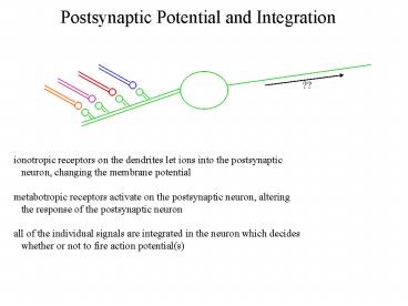
Postsynaptic Potential and Integration
??
ionotropic receptors on the dendrites let ions
into the postsynaptic neuron, changing the
membrane potential metabotropic receptors
activate on the postsynaptic neuron, altering
the response of the postsynaptic neuron all of
the individual signals are integrated in the
neuron which decides whether or not to fire
action potential(s)
2
Stretch Reflex Synaptic Model
the monosynaptic reflex (one excitatory CNS
connection) 1) sensory neurons detect the
stretch of the ligament and muscle 2) dorsal root
ganglia (DRG) signal 2 different types of spinal
neurons a) extensor motor neuron signals to
the muscle, releasing ACh nAChRs on the muscle
open, causing extensor muscle contraction b)
inhibitory interneurons in spinal cord are
activated interneurons block the flexor
motorneurons from firing
3
Stretch Reflex Synaptic Model
artificially depolarizing the sensory neuron
can mimic the reflex causes an EPSP (excitatory
post-synaptic potential) to depolarize the
extensor motor neuron AND interneuron the
interneuron generates an IPSP (inhibitory
postsynaptic potential) in the CNS, 1 synapse
causes approx. a 1 mV postsynaptic voltage
change at the motor end plate, causes a 50 mV
depolarization (1 action potential for one
contraction)
4
Measuring Single Channels
outside-out patch piece of membrane on a patch
pipette with the normal outside of the cell
exposed to the outer solution
single ionotropic receptors 'chatter'- open and
close in rapid succession nAChR's open in an
all or none fashion multiple channels average
together for a more constant current current
amount depends upon the amount of activation
5
Measuring Single Channels
each individual opening lasts for a random period
of time averaging many single channel openings
OR many at once, they have an average open
time can be modeled as a multi-step process with
unbound receptors, singly and doubly bound
open receptors, and bound, closed receptors some
ligand gated channels can even see different
channel openings
6
Measuring Single Channels
nAChRs conduct according to how depolarized the
cell is open times are constant, but more ions
flow through current transferred is directly
proportional to voltage difference no
rectification reversal potential is where
channels may open but no current
flows changing ions on the outside determines
the permeability of the channel to that ion
7
Integrating Single Channels
1 synaptic vesicle can activate many
receptors since conductances add, the total
current I g P n g single channel
conductance modified by potential g (Vm-Vr)
g P probability of a single channel opening
n number of channels opening however,
several things will weaken the synaptic current
membrane capacitance is charge buildup across
the membrane leak currents (pumps and/or
chattering channels) electrical circuits can
model all the conductances comes out to
approximately 1 mV for a normal EPSP in the CNS
8
Integrating Single Channels
action potential repolarization by voltage gated
potassium channels are a rectifier current--
opened only when very depolarized some channels,
like the NMDA receptor, also have rectifying
currents NMDA receptors conduct linearly above a
certain threshold ( -30mV)
9
Inhibitory Postsynaptic Potentials
total current I g P n inhibitory currents
reduce the probability of synaptic
release ie. stimulating inhibitory neurons
causes their targets to be inhibited benzodiaz
apines potentiate GABAA inhibition, making it
stronger without inhibiting neurons by itself
10
Multicomponent Postsynaptic Potentials
both NMDA type and AMPA type glutamate receptors
are found at the same synapses-- two channels
activated by one neurotransmitter separable
using pharmacology AP-5 blocks NMDA channels
AMPA or GYKI block AMPA channels NMDA receptors
have a much longer open time than AMPA channels
AMPA channels open early and, IF the EPSP is
strong enough, NMDA channels open after the
synapse is depolarized inhibitory currents can
have this as well GABA can have a fast (GABAA)
and slow (GABAB) component
11
Slow PSPs are Mediated by Metabotropic Receptors
all neurotransmitters with ionotropic receptors
also have metabotropic metabotropic receptors
mediate slow currents (ie. seconds-minutes) 2
major mechanisms coupling metabotropic receptors
to channels a) direct G-protein coupling
b) indirect coupling through second messengers
12
Slow PSPs are Mediated by Metabotropic Receptors
the duration of a PSP depends upon the rate of
neurotransmitter degradation-- quickly
degraded transmitters do not last for
minutes peptides are not actively degraded, so
may mediate longer currents metabotropic
receptors may also be activated further away from
the synaptic cleft-- changes in the cell
biochemistry can be broader the interneuron
here can cause a strong desensitization
(habituation) to block gill withdrawal when it
is strongly stimulated
13
Slow PSPs are Mediated by Metabotropic Receptors
second messengers can act transcriptionally to
regulate neuronal biochemistry over days to ???
14
Temporal and Spatial Summation
temporal summation occurs when two PSPs occur at
similar times so that the postsynaptic cell
cannot repolarize between potentials
works best where 1 PSP only has a small
volume to change (ie. farther out along a
dendrite) very important for trains of action
potentials
15
Temporal and Spatial Summation
spatial summation requires more than 2 input
neurons
currents can add to one another as ions
diffuse through the dendritic tree will also
always have a time dependent function as well
synapses near the cell body (soma) have a much
stronger effect than those far out along a
dendrite
Recommended
CrystalGraphics Presentations


