Presentaci
1 / 1
Title: Presentaci
1
Effects of repetitive transcranial magnetic
stimulation (rTMS) evaluated by mean of EEG in
the anaesthetized cat Rivadulla C., De Labra C.,
Fernández Del Olmo M. Cudeiro J. NEUROcom,
University of Coruña, Spain.
NEUROcom
RESULTS
INTRODUCTION
rTMS is a non-invasive method that can induce
changes in excitability of the human cortex.
Stimulation at a frequency of around 1Hz
(low-frequency) results in a decrease in
excitability. Conversely, rTMS at frequency of 10
Hz and above (high-frequency) produces the
opposite effect. The brains electrical activity
related with the TMS pulse can be detected with
EEG. Since we used rTMS as a tool to alter visual
cortex activity, we felt it was important to
locate the effect elicited by TMS, and its
spread. Our results show that rTMS effects are
not confined to the vicinity of the stimulation
area and a spread to other regions is observed.
Low frequency rTMS increases low EEG frequencies
and decreases EEG activation induced by visual
stimulation. High-frequency rTMS gave rise to a
different change in the EEG, increasing high
frequencies detected in the FFT power spectrum.
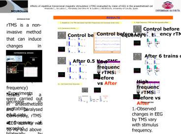
2. High frequency rTMS has the opposite effect
- Repetitive 1 Hz TMS decreases high EEG
frequencies and increases lower ones.
a) On Spontaneous Activity......
EXPERIMENTAL DESIGN
TMS
Visual Stimulation
LGN Extracellular recordings
EEG recordings
b)... And Visual Driven Responses
- Experiments were carried out in anaesthetized and
paralysed adult cats. - EEG activity was recorded bilaterally by mean of
surface electrodes placed on the skull at
frontal, temporal and occipital areas. - rTMS was delivered at low (0.5-1Hz) and high
frequency (15Hz), with a MagStim rapid
stimulator via a 25 mm figure-eight coil, centred
on the occipital lobe. - Activity recorded with and without visual
stimulation in control condition was compared
with the activity recorded in similar conditions
during rTMS. - Calculation of the fast Fourier transform (FFT)
of EEG activity was used to obtain the power
spectrum of data sequences for periods of 1-5 sec
immediately before and after rTMS.


