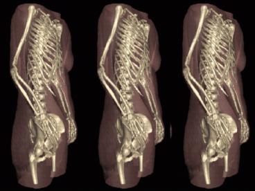
Human AnatomyBio22 - PowerPoint PPT Presentation
1 / 30
Title:
Human AnatomyBio22
Description:
skeleton bones of the skull, vertebral column, and rib cage ... Tubular shaft that forms the axis of long bones ... Structure of Short, Flat, Irregular Bones ... – PowerPoint PPT presentation
Number of Views:95
Avg rating:3.0/5.0
Title: Human AnatomyBio22
1
(No Transcript)
2
Human Anatomy-Bio22 Lecture 6 Bones and Skeletal
Tissues Presented by Tealia Davis, MSc 9-20-05
3
Skeletal Cartilage
- Contains no or
- Surrounded by the (dense irregular
connective tissue) that resists outward expansion - Three types
- 1.
- 2.
- 3.
4
Skeletal Cartilages
- Hyaline
- covers the ends of long bones
- connects the ribs to the sternum
- - makes up the larynx and reinforces air
passages - supports the nose
Fibrocartilage Highly compressed with great
tensile strength Contains fibers Found in
menisci of the knee and in intervertebral discs
Elastic Similar to hyaline cartilage but
contains Found in the external ear and the
epiglottis
5
Bones and Cartilages of the Human Body
6
Classification of Bones
skeleton bones of the skull, vertebral
column, and rib cage skeleton bones of the
upper and lower limbs, shoulder, and hip
7
(No Transcript)
8
(No Transcript)
9
(No Transcript)
10
(No Transcript)
11
(No Transcript)
12
Bone Markings Projections-Sites of Muscle and
Ligament Attachment
rounded projection narrow, prominent
ridge of bone large, blunt, irregular
surface narrow ridge of bone small
rounded projection raised area above a
condyle sharp, slender projection any
bony prominence
13
Bone Markings Projections-Projections that Help
Form Joints
bony expansion carried on a narrow neck
smooth, nearly flat articular surface rounded
articular projection armlike bar of bone
14
Bone Markings Depressions and Openings
canal-like passageway cavity within a
bone shallow, basinlike depression
furrow narrow, slitlike opening round or
oval opening through a bone
15
(No Transcript)
16
Structure of Long Bone
Long bones consist of a diaphysis and an
epiphysis Tubular shaft that forms the axis
of long bones Composed of compact bone that
surrounds the medullary cavity, Yellow bone
marrow (fat) is contained in the medullary
cavity Expanded ends of long
bones Exterior is compact bone, and the interior
is spongy bone, Joint surface is covered with
articular (hyaline) cartilage, Epiphyseal line
separates the diaphysis from the epiphyses
17
(No Transcript)
18
(No Transcript)
19
(No Transcript)
20
(No Transcript)
21
Microscopic Structure Compact Bone
Haversian system, or osteon the structural unit
of compact bone Lamella weight-bearing,
column-like matrix tubes composed mainly of
collagen Haversian, or central canal central
channel containing blood vessels and
nerves Volkmanns canals channels lying at
right angles to the central canal, connecting
blood and nerve supply of the periosteum to that
of the Haversian canal Osteocytes mature bone
cells Lacunae small cavities in bone that
contain osteocytes
22
(No Transcript)
23
Components of Bone
- Organic
- bone-forming cells
- mature bone cells
- large cells that reasorb or break down bone
matrix - unmineralized bone matrix composed of
proteoglycans, glycoproteins, and collagen
Inorganic , or mineral salts Sixty-five
percent of bone by mass Mainly calcium
phosphates Responsible for bone hardness and its
resistance to compression
24
Bone Development
Osteogenesis and ossification the process of
bone tissue formation, which leads to The
formation of the bony skeleton in embryos Bone
growth until early adulthood Bone thickness,
remodeling, and repair
Ossification Intramembranous Ossification Endoch
ondral Ossification
25
Postnatal Bone Growth
Growth in length of long bones Cartilage on the
side of the epiphyseal plate closest to the
epiphysis is relatively inactive Cartilage
abutting the shaft of the bone organizes into a
pattern that allows fast, efficient growth
Cells of the epiphyseal plate proximal to the
resting cartilage form three functionally
different zones growth, transformation, and
osteogenic
26
Functional Zones in Bone Growth
Growth zone cartilage cells undergo mitosis,
pushing the epiphysis away from the
diaphysis Transformation zone older cells
enlarge, the matrix becomes calcified, cartilage
cells die, and the matrix begins to
deteriorate Osteogenic zone new bone formation
occurs
27
(No Transcript)
28
(No Transcript)
29
(No Transcript)
30
SEE YOU ON TUESDAY!


